Copyright
©The Author(s) 2022.
World J Clin Cases. May 16, 2022; 10(14): 4640-4647
Published online May 16, 2022. doi: 10.12998/wjcc.v10.i14.4640
Published online May 16, 2022. doi: 10.12998/wjcc.v10.i14.4640
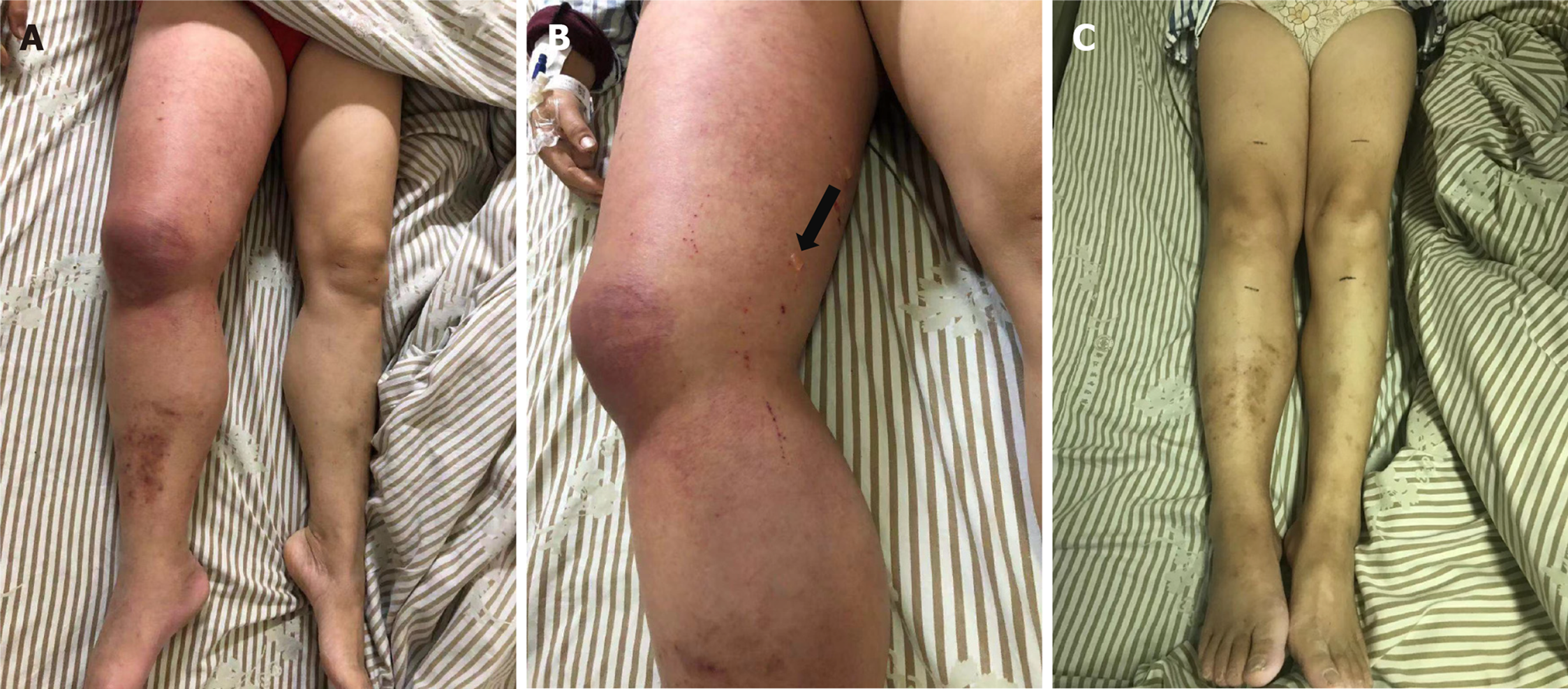
Figure 1 The right lower limb of the patient.
A: The skin of the right lower limb became red; B: Swollen with several soybean-sized transparent blisters before therapy (the black arrow indicates the blister site); C: Both lower limbs of the patient had the same circumference after 3 wk of treatment with fondaparinux sodium.
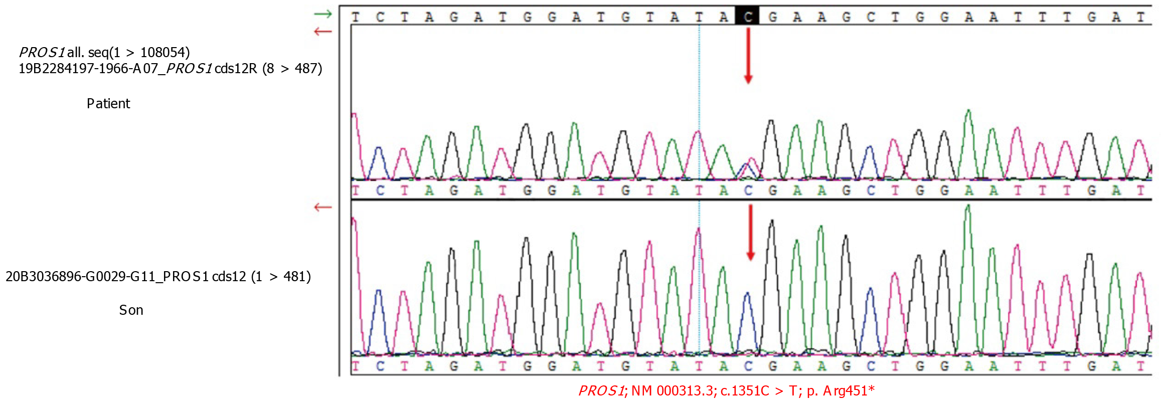
Figure 2 Sanger sequencing results indicating a positive PROS1 mutation [c.
1351C > T (p. Arg451)] (red arrow indicates the mutation site). NM: RefSeq of mRNA.
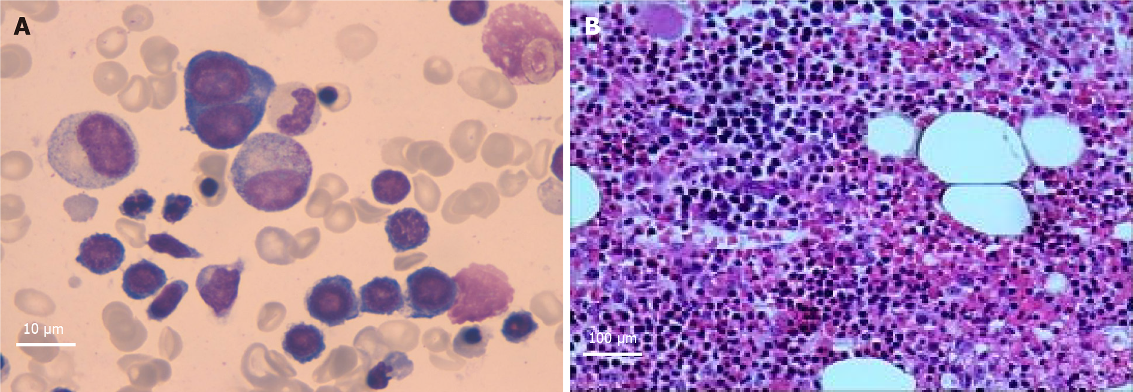
Figure 3 Bone marrow cell examination and bone marrow biopsy.
A: Bone marrow cell examination (Magnification: 100 ×); B: Bone marrow biopsy (Magnification: 10 ×).
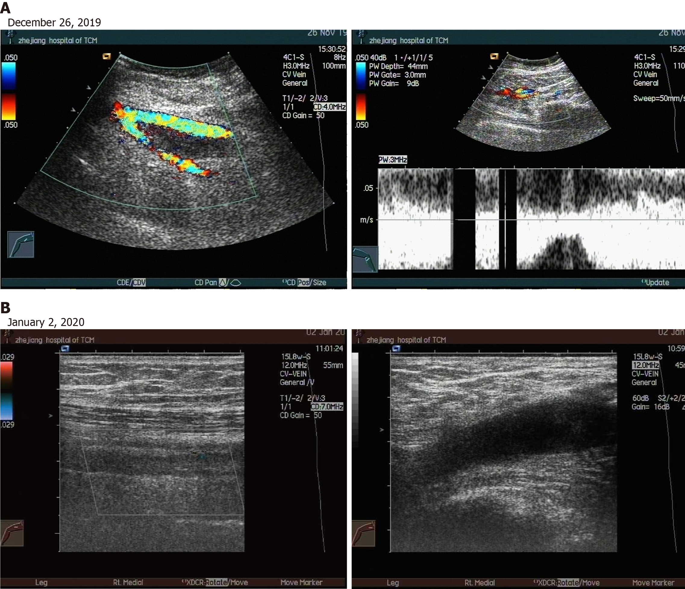
Figure 4 Deep venous ultrasound of the right lower extremity.
A: Lower extremity vascular ultrasound: Extensive acute incomplete thrombosis in the right lower extremities; B: Lower extremity vascular ultrasound: Old thrombosis in the common and superficial femoral veins of the right lower limb.
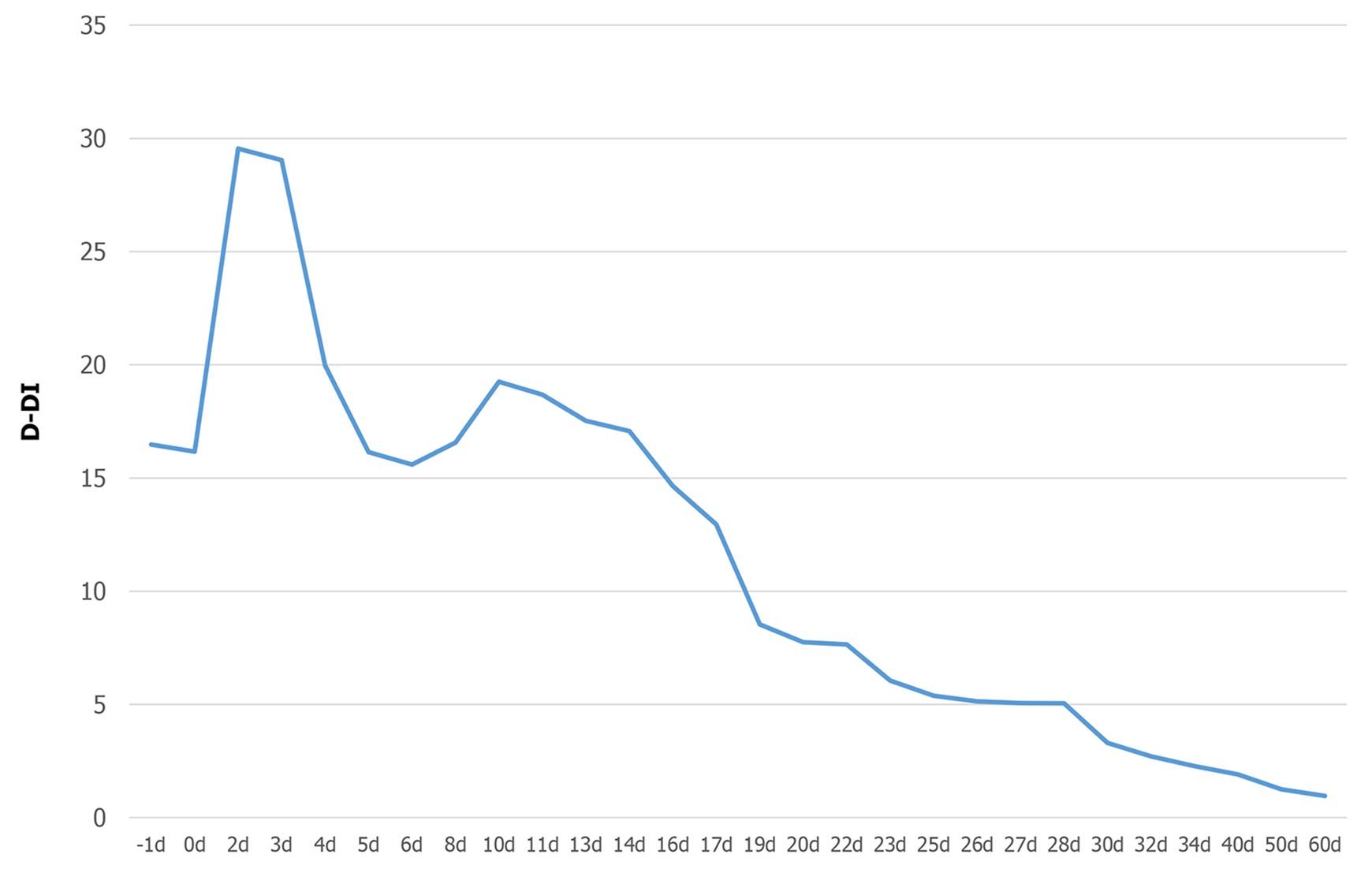
Figure 5 The change in D-dimer during fondaparinux sodium therapy.
D-DI: D-dimer.
- Citation: Liu WB, Ma JX, Tong HX. Successful treatment in one myelodysplastic syndrome patient with primary thrombocytopenia and secondary deep vein thrombosis: A case report . World J Clin Cases 2022; 10(14): 4640-4647
- URL: https://www.wjgnet.com/2307-8960/full/v10/i14/4640.htm
- DOI: https://dx.doi.org/10.12998/wjcc.v10.i14.4640


