Copyright
©The Author(s) 2022.
World J Clin Cases. May 16, 2022; 10(14): 4541-4549
Published online May 16, 2022. doi: 10.12998/wjcc.v10.i14.4541
Published online May 16, 2022. doi: 10.12998/wjcc.v10.i14.4541
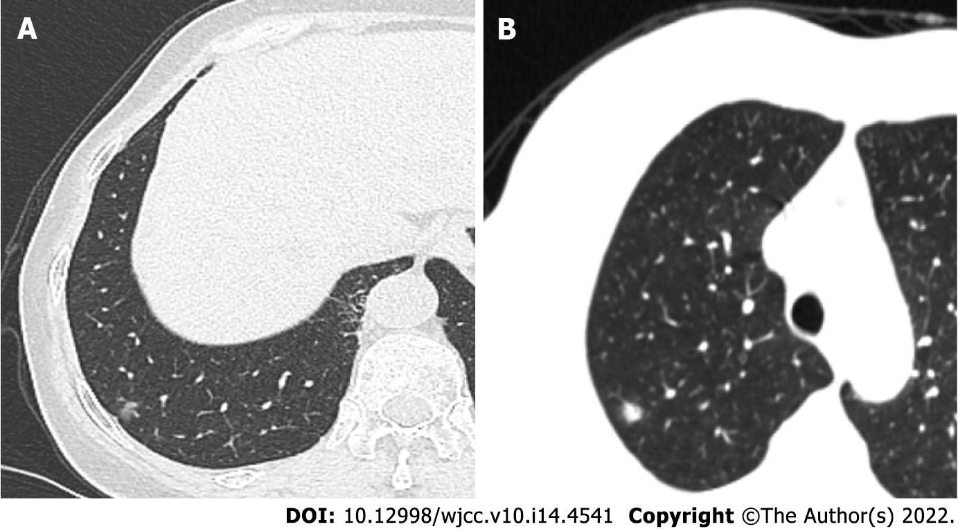
Figure 1 Imaging findings of pulmonary nodules in case 1 and case 2.
A: A mixed ground-glass nodule with a diameter of 0.6 cm in the subpleura of the posterior basal segment of the lower lobe of the right lung in case 1. The texture was relatively uniform, and the nodule was slightly pulled near the pleura; B: A solid lobulated nodule at the apex of the right upper lobe, 8 mm in diameter, with blurred edge, in case 2.
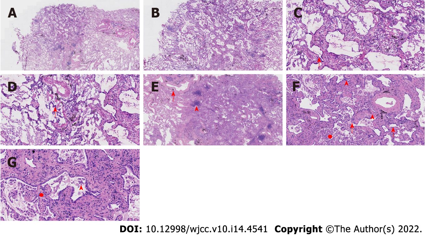
Figure 2 Pathological features of case 1.
A: At low magnification (40 ×, frozen section), the boundary of the tumor was relatively clear, and there was air cavities; B: At low magnification (100 ×, frozen section), the boundary of the tumor was relatively clear, and there was air cavities; C and D: Observations at high magnification (200 ×, frozen section) revealed that the tumor cells were mainly arranged in a monolayer structure, and the local part seemed to be a bilayer structure. Morphologically, the cells were observed to be medium sized, the nuclear chromatin was pale and homogeneous, and local cilia were seen (red arrow); E: At low magnification (100 ×), the relationship between the pulmonary lobular artery and bronchioles was close (arrow), and peripheral stromal lymphocytes were infiltrated in a focal shape (triangle); F and G: Observations at medium to high magnification (200 × and 400 ×, respectively) revealed that tumor cells were arranged as papillary and mural structures. The cell morphology is mild with visible cilia (arrows), bilayer structures (triangles), aggregation of phagocytes in the alveolar cavity (circle, F), and a fibrous non-cancerous stroma (circle, G).
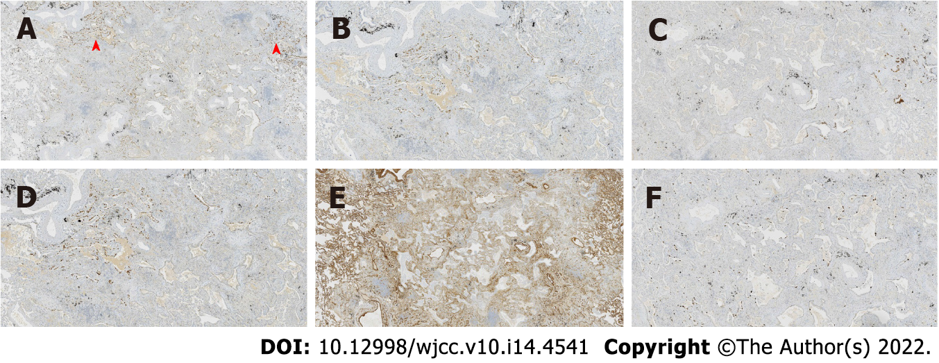
Figure 3 Immunohistochemical staining in case 1.
Thyroid transcription factor 1 was expressed in bronchioles and the surrounding tumor glands, with the only difference being intensity. The results of P40, P63, and cytokeratin 5/6 staining were the same, and positive staining was detected only in the bilayer structures of the tumor. Collagen IV staining showed the presence of alveolar structure, and the Ki-67 index was low. A: Thyroid transcription factor 1; B: P40; C: Cytokeratin 5/6; D: P63; E: Collagen IV; F: Ki-67.
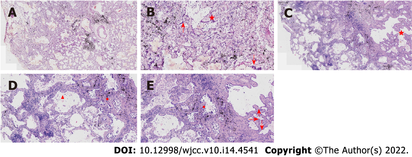
Figure 4 Pathological features of case 2.
A: At low magnification (100 ×, frozen section), the boundary of the tumor was relatively clear; there were air cavities and arterioles were visible; B: At high magnification (200 ×, frozen section), the tumor cells were found to be mainly arranged in a monolayer with locally visible cilia (arrows); some nuclei appeared enlarged and atypical (star); C: At low magnification (100 ×), most cells appeared with moderate density (star), focal hyperplasia, and stroma within the focal lymphocytic infiltration; D and E: Observations at medium to high magnification (200 × and 400 ×, respectively) revealed that the tumor cells were arranged as an acinar structure and accessory wall structure; most cells were not atypia in shape, and cilia (arrow) were seen. Some nuclei were enlarged and atypical (circle).
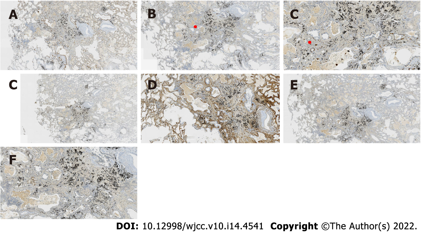
Figure 5 Immunohistochemical staining in case 2.
Thyroid transcription factor 1 was positive; the results for P63 and CK5/6 staining were the same, and only basal cells were shown in the hyperplasia area. CD34 showed the presence of alveolar structure, and the Ki-67 index was low. A: Thyroid transcription factor 1; B: P63; C: Cytokeratin 5/6; D: CD34; E: Ki-67; F: P53.
- Citation: Du Y, Wang ZY, Zheng Z, Li YX, Wang XY, Du R. Bronchiolar adenoma with unusual presentation: Two case reports. World J Clin Cases 2022; 10(14): 4541-4549
- URL: https://www.wjgnet.com/2307-8960/full/v10/i14/4541.htm
- DOI: https://dx.doi.org/10.12998/wjcc.v10.i14.4541


