Copyright
©The Author(s) 2022.
World J Clin Cases. May 6, 2022; 10(13): 4190-4195
Published online May 6, 2022. doi: 10.12998/wjcc.v10.i13.4190
Published online May 6, 2022. doi: 10.12998/wjcc.v10.i13.4190
Figure 1 Paraneoplastic antibodies.
A, B: Paraneoplastic antibodies in cerebrospinal fluid on July 20, 2020 (Fluorescent picture, monkey cerebellum); C, D: Paraneoplastic antibodies in blood on July 20, 2020 (Fluorescent picture, monkey cerebellum).
Figure 2 Computed tomography findings on July 18, 2020.
A-C: The bronchial lumen in the lower lobe of the right lung was unevenly narrowed, and irregular nodular shadow of about 4.6 cm × 3.6 cm was seen, and the superficial lobar sign was visible
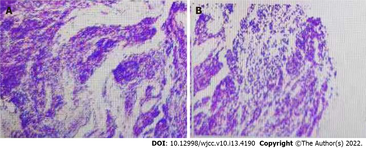
Figure 3 Bronchial biopsy pathology report on July 30, 2020.
A, B: The bronchial biopsy pathology report showed small cell lung cancer.
- Citation: Li ZC, Cai HB, Fan ZZ, Zhai XB, Ge ZM. Paraneoplastic neurological syndrome with positive anti-Hu and anti-Yo antibodies: A case report. World J Clin Cases 2022; 10(13): 4190-4195
- URL: https://www.wjgnet.com/2307-8960/full/v10/i13/4190.htm
- DOI: https://dx.doi.org/10.12998/wjcc.v10.i13.4190


