Copyright
©The Author(s) 2022.
World J Clin Cases. May 6, 2022; 10(13): 4145-4152
Published online May 6, 2022. doi: 10.12998/wjcc.v10.i13.4145
Published online May 6, 2022. doi: 10.12998/wjcc.v10.i13.4145
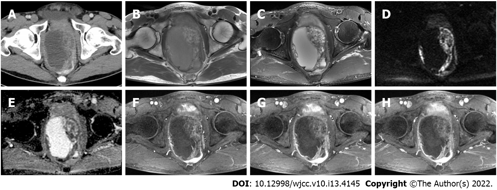
Figure 1 Preoperative pelvic imaging.
A: Contrast-enhanced computed tomography shows a large, cystic-solid mass in the pelvis; B and C: At magnetic resonance imaging (MRI), the lesion near the prostate is isointense to slightly hyperintense on T1-weighted imaging (WI), with heterogeneous intensity on T2WI; D and E: There is heterogeneous hyperintensity on diffusion-WI with opposing hypointensity on the apparent diffusion coefficient maps; F-H: The solid part is enhanced during the arterial phase on contrast-enhanced MRI, with persistent enhancement in the venous and delayed phases. No enhancement is seen in the cystic part.
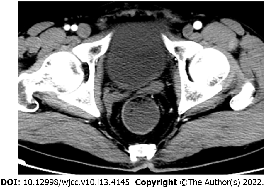
Figure 2 Post-operative contrast-enhanced pelvic computed tomography.
Imaging after the first cycle of chemotherapy at 2 mo postoperatively shows no obvious signs of residual tumor or recurrence.
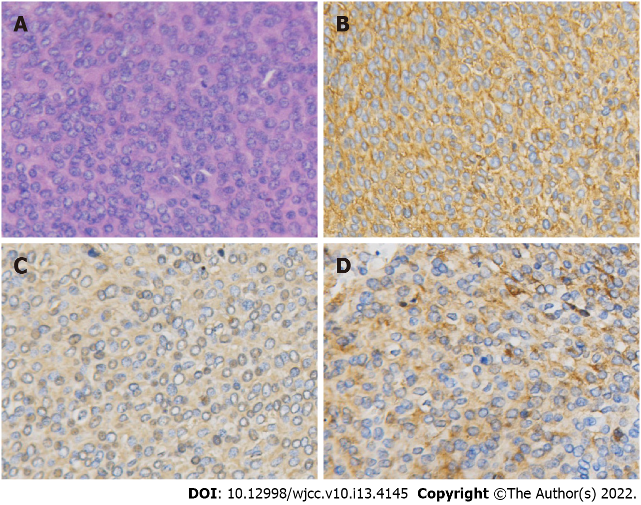
Figure 3 Hematoxylin-eosin and immunohistochemical staining.
A: Hematoxylin-eosin staining reveals small round cells arranged closely in a flaky pattern (× 200); B: Immunohistochemical staining of the small round cells for CD99 shows strong positivity for this marker (× 200); C and D: Immunohistochemical staining for vimentin and synaptophysin is positive (× 200).
- Citation: Tian DW, Wang XC, Zhang H, Tan Y. Primitive neuroectodermal tumor of the prostate in a 58-year-old man: A case report. World J Clin Cases 2022; 10(13): 4145-4152
- URL: https://www.wjgnet.com/2307-8960/full/v10/i13/4145.htm
- DOI: https://dx.doi.org/10.12998/wjcc.v10.i13.4145


