Copyright
©The Author(s) 2022.
World J Clin Cases. May 6, 2022; 10(13): 4020-4032
Published online May 6, 2022. doi: 10.12998/wjcc.v10.i13.4020
Published online May 6, 2022. doi: 10.12998/wjcc.v10.i13.4020
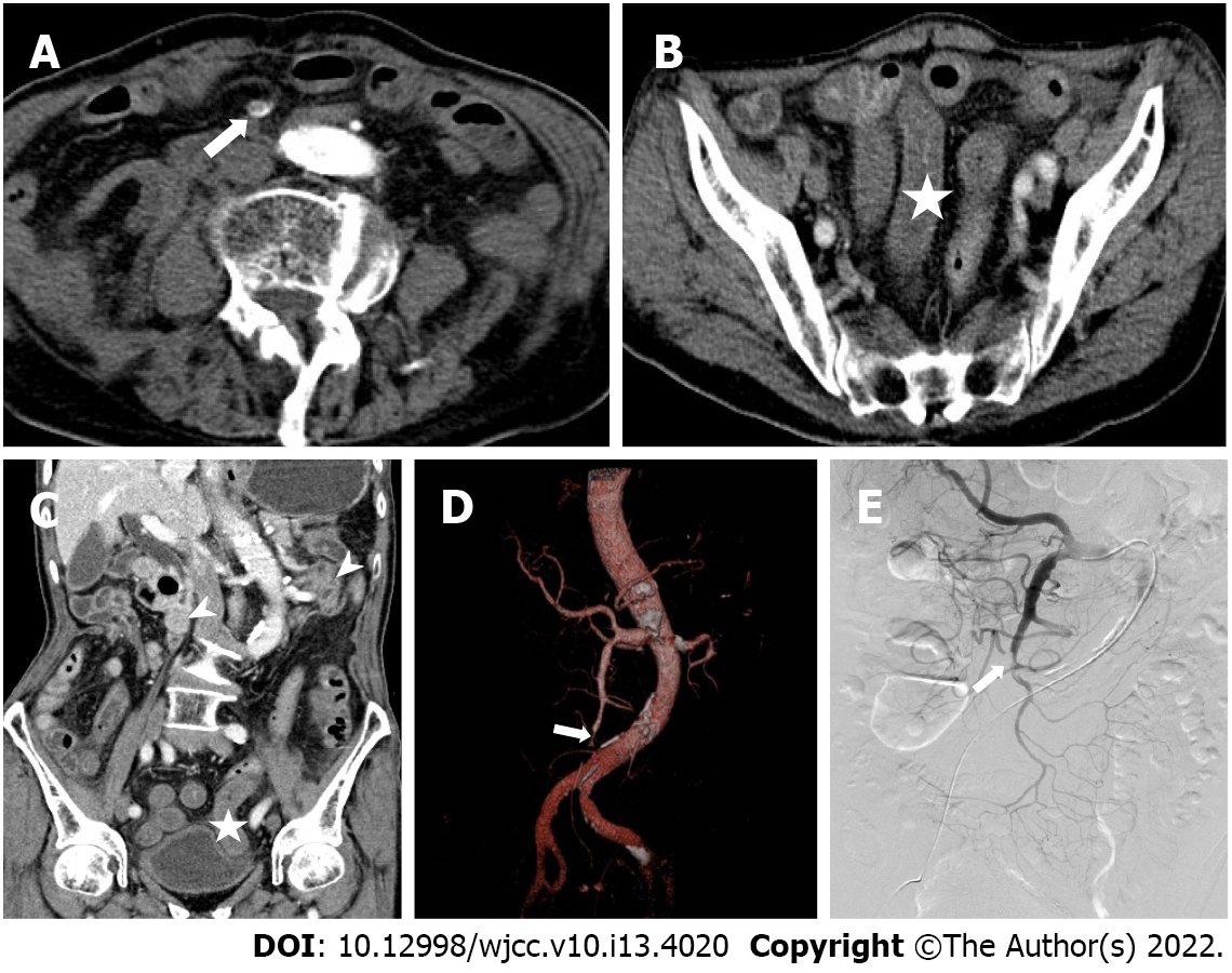
Figure 1 An 89 years old male with sudden severe abdominal pain was hospitalized for 1 d.
A: The axial image of arterial phase on computed tomography enhanced scan, showing diffuse embolism (long arrow) in superior mesenteric artery III and IV regions; B: An axial image of venous phase, showing thickening of intestinal wall and decreased enhancement (asterisk) at the end of ileum; C: The coronal image of venous phase. It can be seen that the enhancement of ileum is significantly lower than that of normal intestinal wall (arrow); D and E: Volume rendered technique and digital subtraction images, respectively, the proximal ileocolic artery and ileal artery are not displayed.
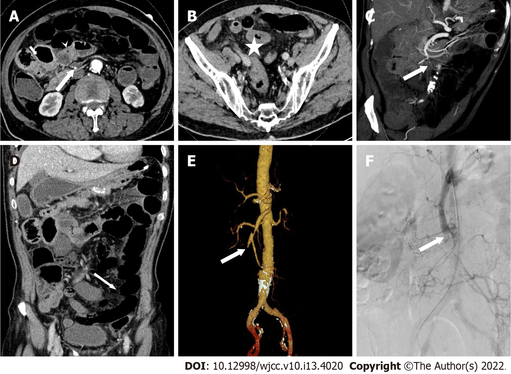
Figure 2 Male, 71 years old, abdominal pain for 18 h.
A: The axial image of enhanced computed tomography in arterial phase; B and D: The axial and coronal images in venous phase; C: The oblique coronal maximum intensity projection image; E: Volume rendered technique; and F: Digital subtraction. It showed diffuse embolism (long arrow) in superior mesenteric artery III and IV regions, accompanied by intestinal wall thickening (asterisk), intestinal wall thinning (slender arrow), decreased enhancement relative to normal intestinal wall (arrow), intestinal cavity expansion and mesenteric fat stranding. Imaging diagnosis of extensive ischemia of small intestine.
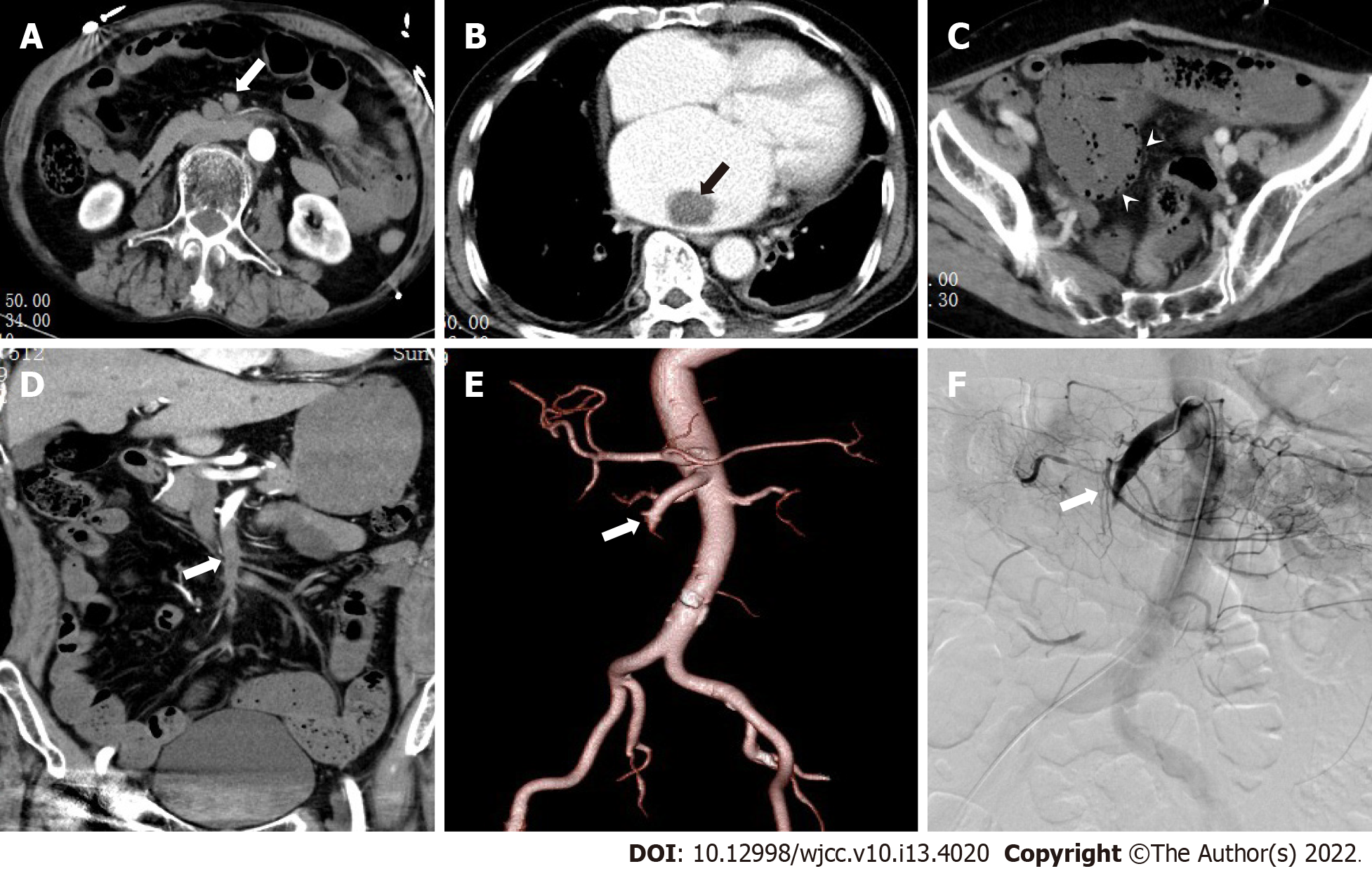
Figure 3 The patient was a 62-year-old male with history of abdominal pain, hematochezia, and atrial fibrillation.
A and B: The axial images in the arterial phase; C: The axial images in the venous phase; D: The coronary images in the arterial phase; E: The volume rendered technique image; and F: The digital subtraction image. A and D show diffuse embolism in II, III, and IV regions of superior mesenteric artery (long arrows); B shows embolus in the left atrium, C shows decreased intestinal wall enhancement, and signs of pneumatosis intestinalis (arrow).
- Citation: Yang JS, Xu ZY, Chen FX, Wang MR, Cong RC, Fan XL, He BS, Xing W. Role of clinical data and multidetector computed tomography findings in acute superior mesenteric artery embolism. World J Clin Cases 2022; 10(13): 4020-4032
- URL: https://www.wjgnet.com/2307-8960/full/v10/i13/4020.htm
- DOI: https://dx.doi.org/10.12998/wjcc.v10.i13.4020


