Copyright
©The Author(s) 2022.
World J Clin Cases. Apr 26, 2022; 10(12): 3930-3935
Published online Apr 26, 2022. doi: 10.12998/wjcc.v10.i12.3930
Published online Apr 26, 2022. doi: 10.12998/wjcc.v10.i12.3930
Figure 1 Diffuse autoimmune pancreatitis complicated by gastric varices.
A: Computed tomography showed a diffusely enlarged pancreas with a capsule-like rim,obstructed splenic vein and slight splenomegaly; B: Magnetic resonance cholangiopancreatography showed the irregular expanding of pancreatic duct in the neck and body of pancreas, and the swelling of pancreas; C: Esophagogastroduodenoscopy showed the partial gastric varices in fundus with positive red-color sign.
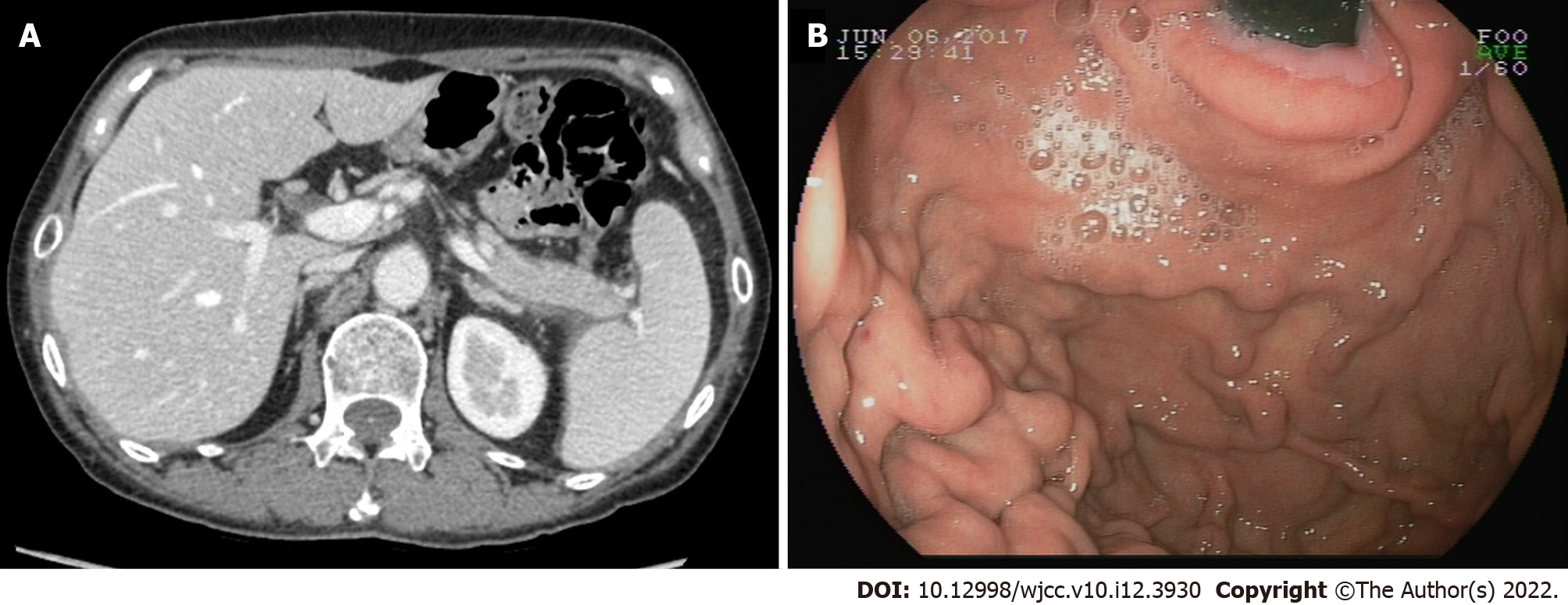
Figure 2 Autoimmune pancreatitis was significantly improved after two month steroid therapy.
A and B: Computed tomography and esophagogastroduodenoscopy showed the improvement of swelling pancreas,obstructed splenic vein, splenomegaly (A) and gastric varices with positive red-color sign (B).
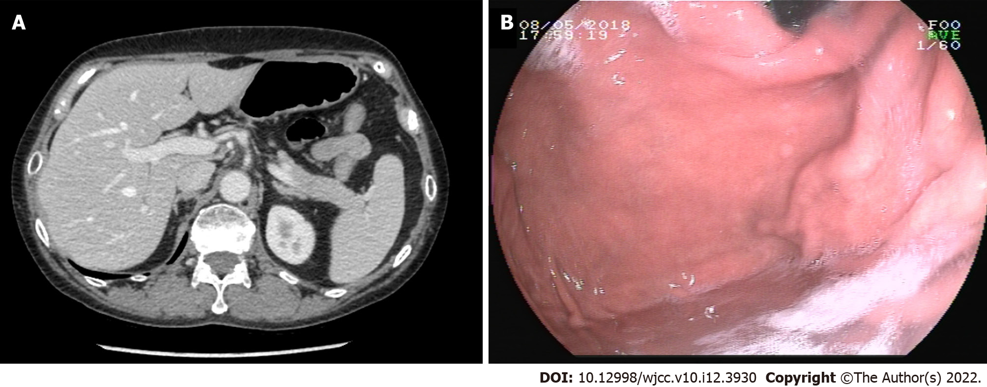
Figure 3 Autoimmune pancreatitis was further improved after five month steroid therapy.
A and B: Computed tomography and esophagogastroduodenoscopy showed the improvement of swelling pancreas, obstructed splenic vein, splenomegaly (A) and gastric varices with negative red-color sign (B).
Figure 4 Gastric varices disappeared after 1 year steroid therapy.
A: Computed tomography showed the pancreas, spleen vein and spleen restore to the normal level; B: Esophagogastroduodenoscopy showed the gastric varices disappeared.
- Citation: Hao NB, Li X, Hu WW, Zhang D, Xie J, Wang XL, Li CZ. Steriod for Autoimmune pancreatitis complicating by gastric varices: A case report. World J Clin Cases 2022; 10(12): 3930-3935
- URL: https://www.wjgnet.com/2307-8960/full/v10/i12/3930.htm
- DOI: https://dx.doi.org/10.12998/wjcc.v10.i12.3930


