Copyright
©The Author(s) 2022.
World J Clin Cases. Apr 26, 2022; 10(12): 3916-3922
Published online Apr 26, 2022. doi: 10.12998/wjcc.v10.i12.3916
Published online Apr 26, 2022. doi: 10.12998/wjcc.v10.i12.3916
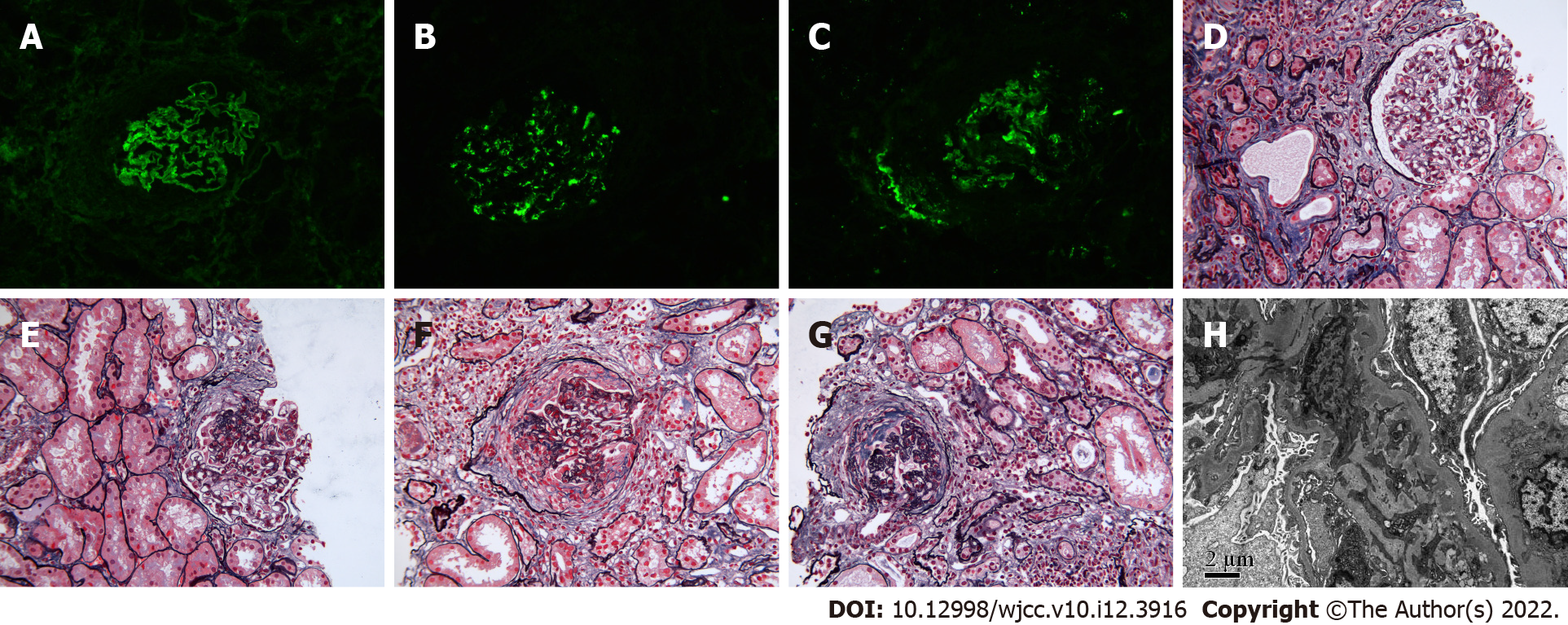
Figure 1 Representative histopathology pictures of concurrent anti-glomerular basement membrane disease and Immunoglobulin A nephropathy.
A: Immunofluorescence analysis showed strong (3+) staining of IgG along the linear capillary loop (original magnification, × 200); B: Immunofluorescence analysis showed strong (3+) Immunoglobulin A (IgA) staining indicating granular and bolus-type depositions of IgA in the mesangial area (original magnification, × 200); C: Immunofluorescence analysis showed strong (3+) C3 staining indicating granular and bolus-type depositions of C3 in the mesangial area (original magnification, × 200; D: Light microscopy analysis showed small cell crescents (photoacoustic shadow-casting microscopy, × 200); E: Light microscopy showed a small cellular fibrous crescent (photoacoustic shadow-casting microscopy, × 200; F: Light microscopy analysis showed cellular fibrous crescents (photoacoustic shadow-casting microscopy, × 200); G: Light microscopy analysis showed fibrous crescents with sclerosis (photoacoustic shadow-casting microscopy, × 200; H: Electron microscopy analysis showed massive electron-dense deposits in the mesangial area (original magnification, × 8000).
- Citation: Guo C, Ye M, Li S, Zhu TT, Rao XR. Anti-glomerular basement membrane disease with IgA nephropathy: A case report. World J Clin Cases 2022; 10(12): 3916-3922
- URL: https://www.wjgnet.com/2307-8960/full/v10/i12/3916.htm
- DOI: https://dx.doi.org/10.12998/wjcc.v10.i12.3916


