Copyright
©The Author(s) 2022.
World J Clin Cases. Apr 16, 2022; 10(11): 3541-3546
Published online Apr 16, 2022. doi: 10.12998/wjcc.v10.i11.3541
Published online Apr 16, 2022. doi: 10.12998/wjcc.v10.i11.3541
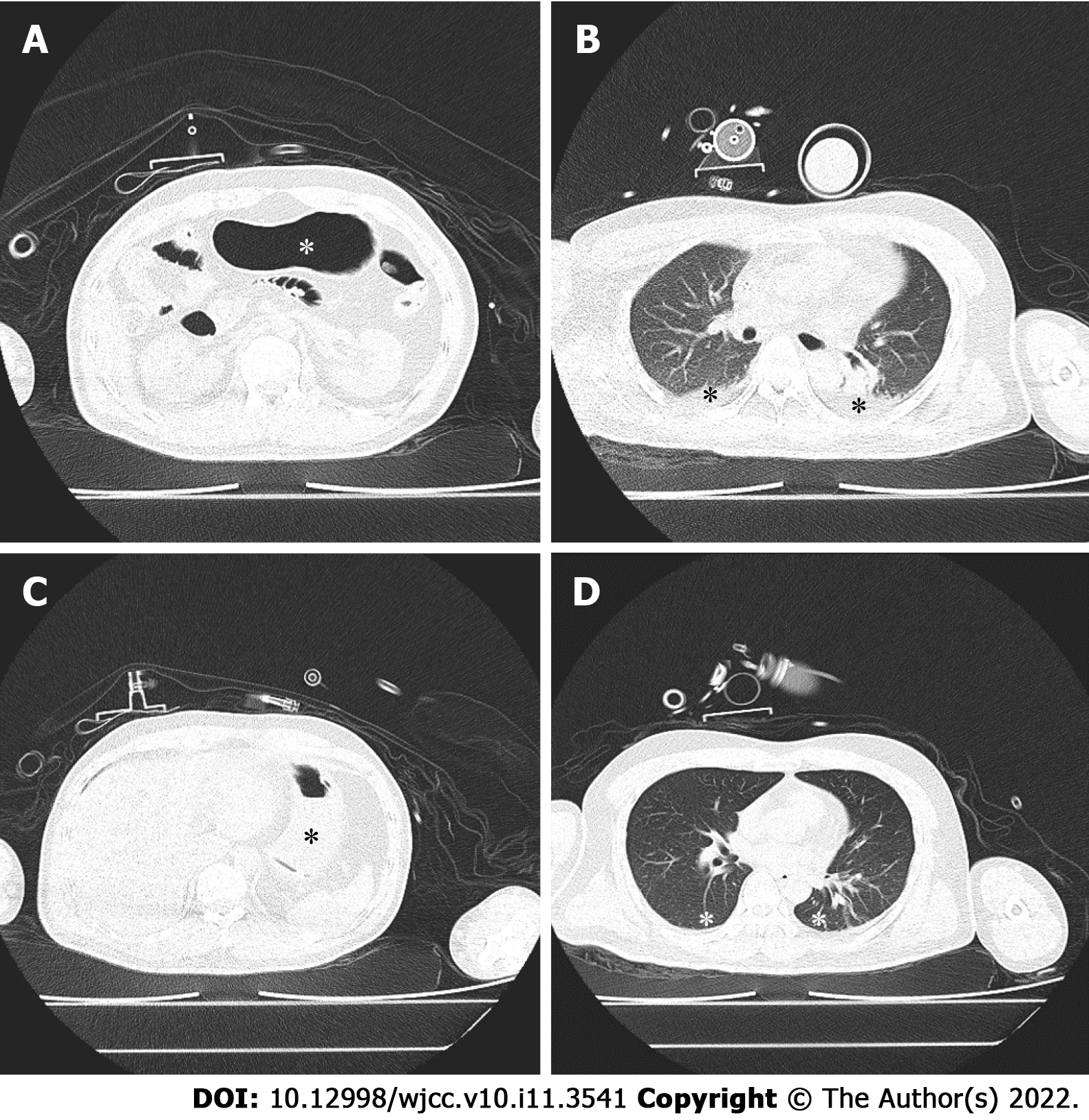
Figure 1 Patient’s computed tomography of upper abdomen (A and C) and lung (B and D).
Before gastric decompression, gastric insufflation (A, asterisk) and atelectasis with air bronchogram (B, asterisk) could be seen. After gastric decompression and standard lung recruitment manoeuvre, the stomach was deflated (C, asterisk) and atelectatic lung tissue (D, asterisk) was re-aerated.
- Citation: Zhao Y, Li P, Li DW, Zhao GF, Li XY. Severe gastric insufflation and consequent atelectasis caused by gas leakage using AIR-Q laryngeal mask airway: A case report. World J Clin Cases 2022; 10(11): 3541-3546
- URL: https://www.wjgnet.com/2307-8960/full/v10/i11/3541.htm
- DOI: https://dx.doi.org/10.12998/wjcc.v10.i11.3541


