Copyright
©The Author(s) 2022.
World J Clin Cases. Apr 16, 2022; 10(11): 3533-3540
Published online Apr 16, 2022. doi: 10.12998/wjcc.v10.i11.3533
Published online Apr 16, 2022. doi: 10.12998/wjcc.v10.i11.3533
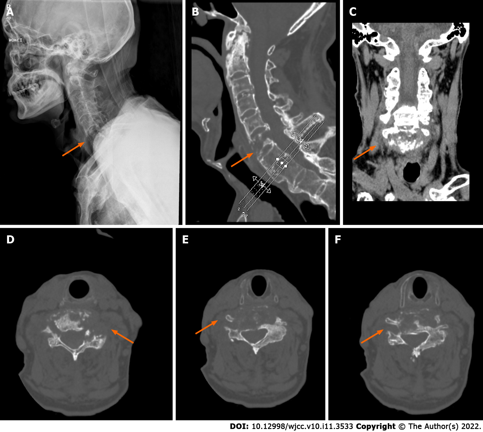
Figure 1 Preoperative X-ray and computed tomography.
A: Lateral X-ray view, an arrow shows pathological fracture; B: CT sagittal image, an arrow shows pathological fracture; C: Computed tomography (CT) coronal image, an arrow shows pathological fracture; D-F: CT axial images at different slice levels, an arrow shows pathological fracture.
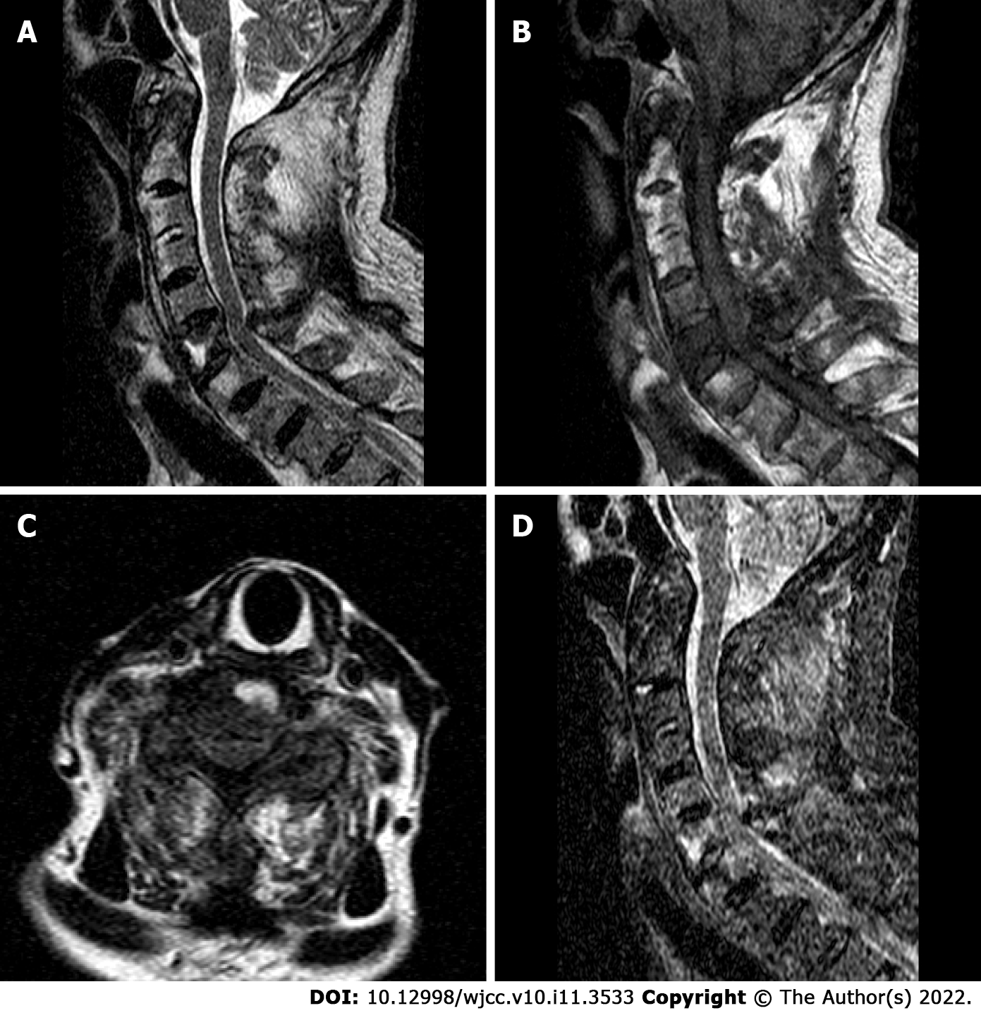
Figure 2 Preoperative magnetic resonance imaging.
A: Sagittal T2-weighted image; B: Sagittal T1-weighted image; C: Axial T2-weighted image; D: Sagittal STIR image.
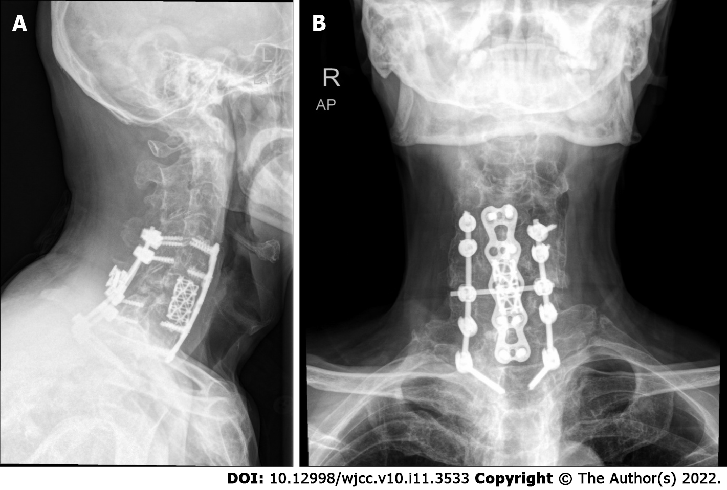
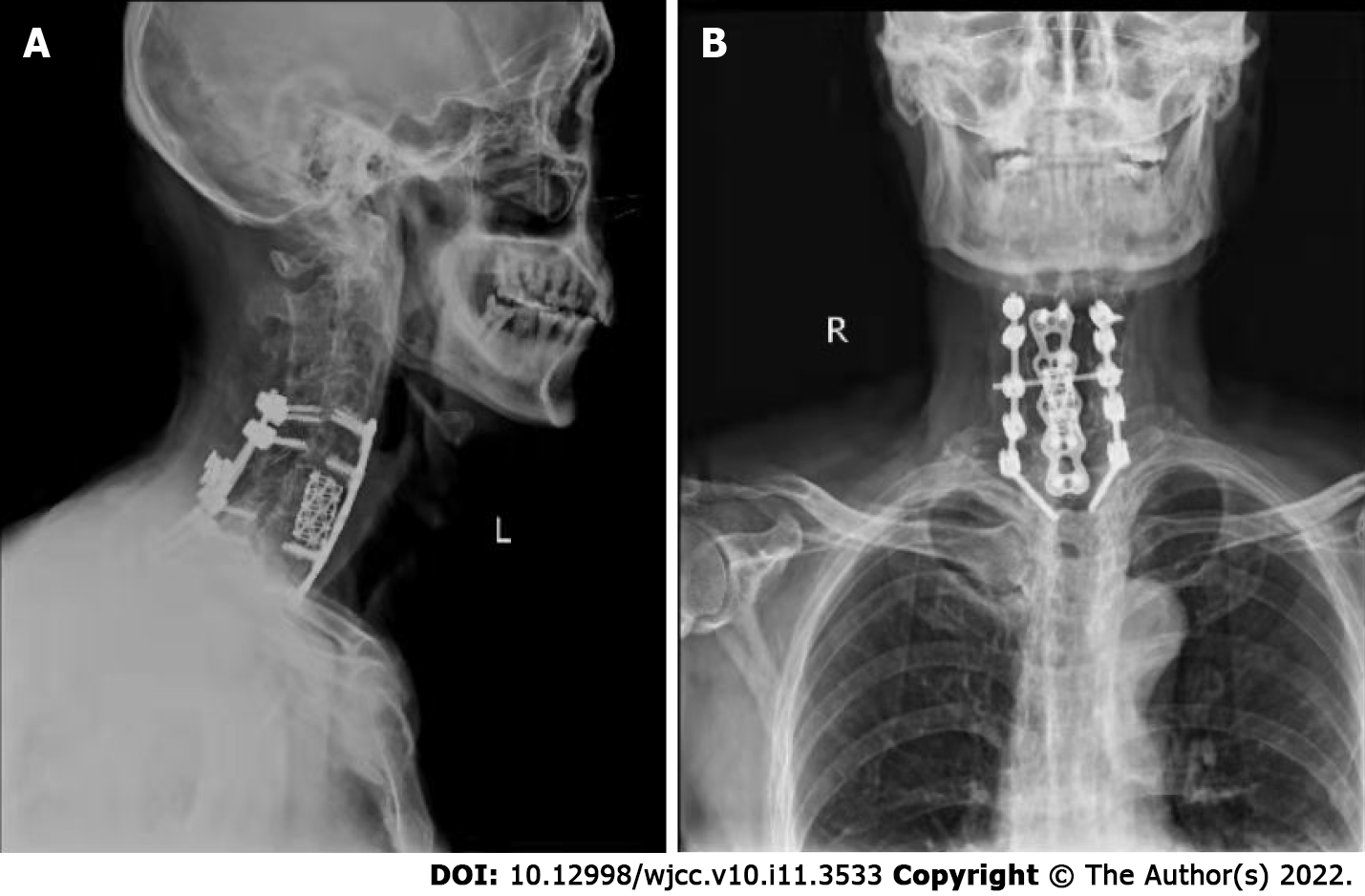
Figure 3 Postoperative X-ray.
A: Lateral X-ray view; B: Anteroposterior X-ray view.
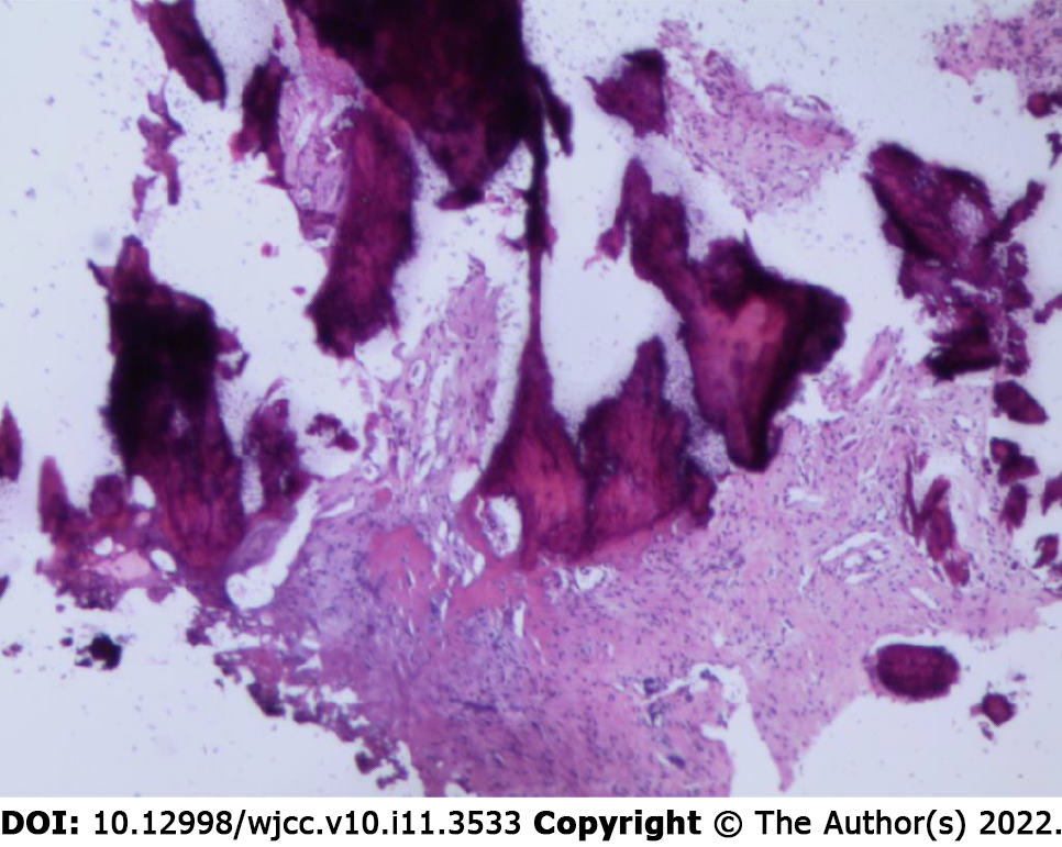
Figure 4 Pathology picture.
Figure 5 10 months postoperative X-ray.
A: Lateral X-ray view; B: Anteroposterior X-ray view.
- Citation: Peng YJ, Zhou Z, Wang QL, Liu XF, Yan J. Ankylosing spondylitis complicated with andersson lesion in the lower cervical spine: A case report. World J Clin Cases 2022; 10(11): 3533-3540
- URL: https://www.wjgnet.com/2307-8960/full/v10/i11/3533.htm
- DOI: https://dx.doi.org/10.12998/wjcc.v10.i11.3533


