Copyright
©The Author(s) 2022.
World J Clin Cases. Apr 16, 2022; 10(11): 3527-3532
Published online Apr 16, 2022. doi: 10.12998/wjcc.v10.i11.3527
Published online Apr 16, 2022. doi: 10.12998/wjcc.v10.i11.3527
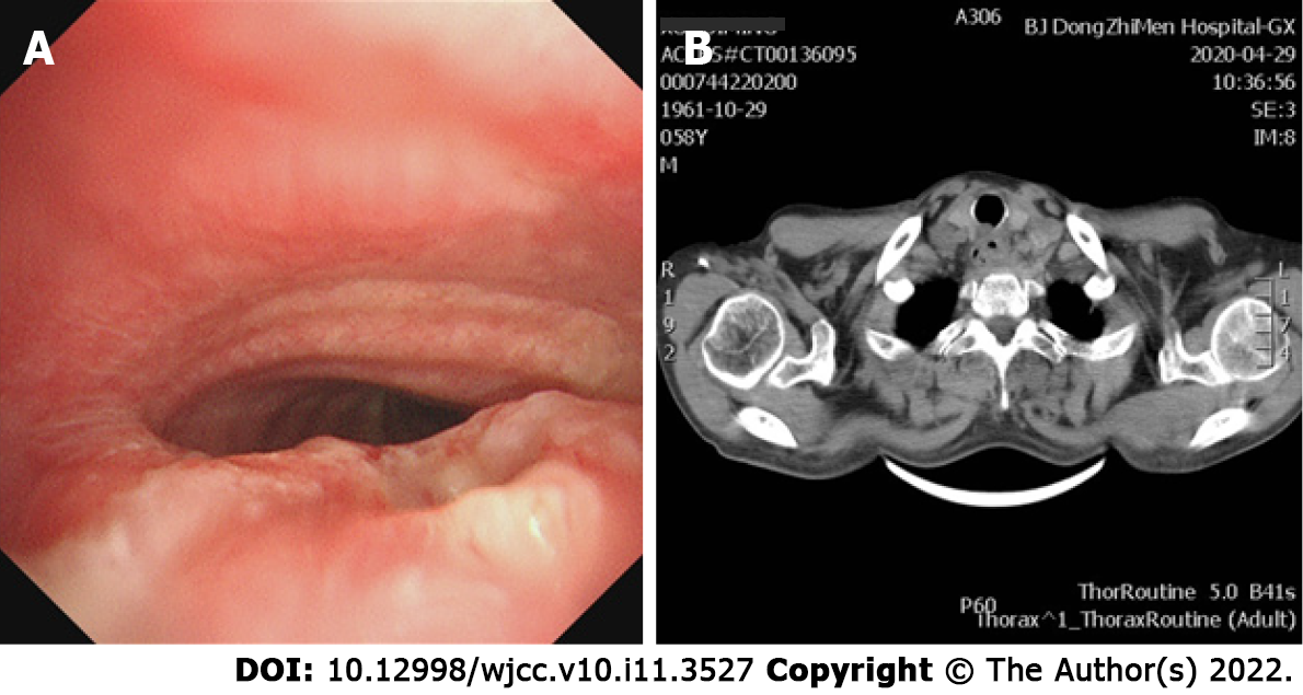
Figure 1 Tracheoesophageal fistula and narrowing of the main bronchus.
A: Bronchoscopy image; B: Computed tomography image.
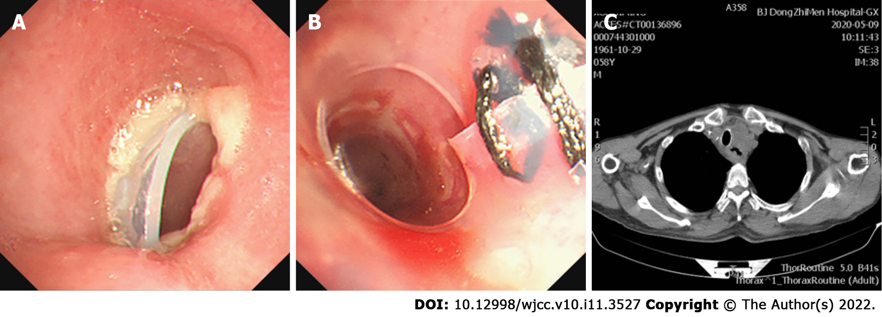
Figure 2 Fistula completely enclosed by a Y-shaped and modified straight silicone stent placed in the main trachea.
A, B: Bronchoscopy image showing (A) the upper edge of Y-shaped silicone stent incarcerated the airway wall and (B) a Y-shaped silicone stent and a straight silicone stent placed after mechanical debulking of the tumor; C: Computed tomography image of B.
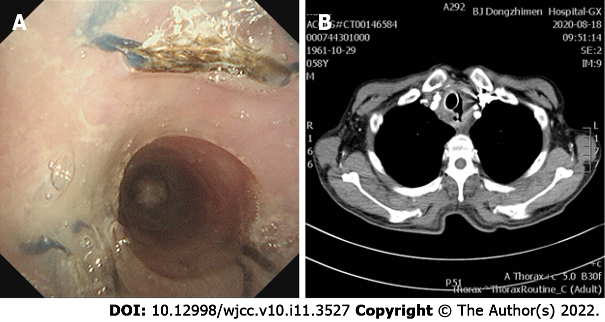
Figure 3 Original malignant tracheoesophageal fistula following rapid progression after the fourth dose of toripalimab.
A: Bronchoscopy image; B: Computed tomography image showing the fistula having progressed.
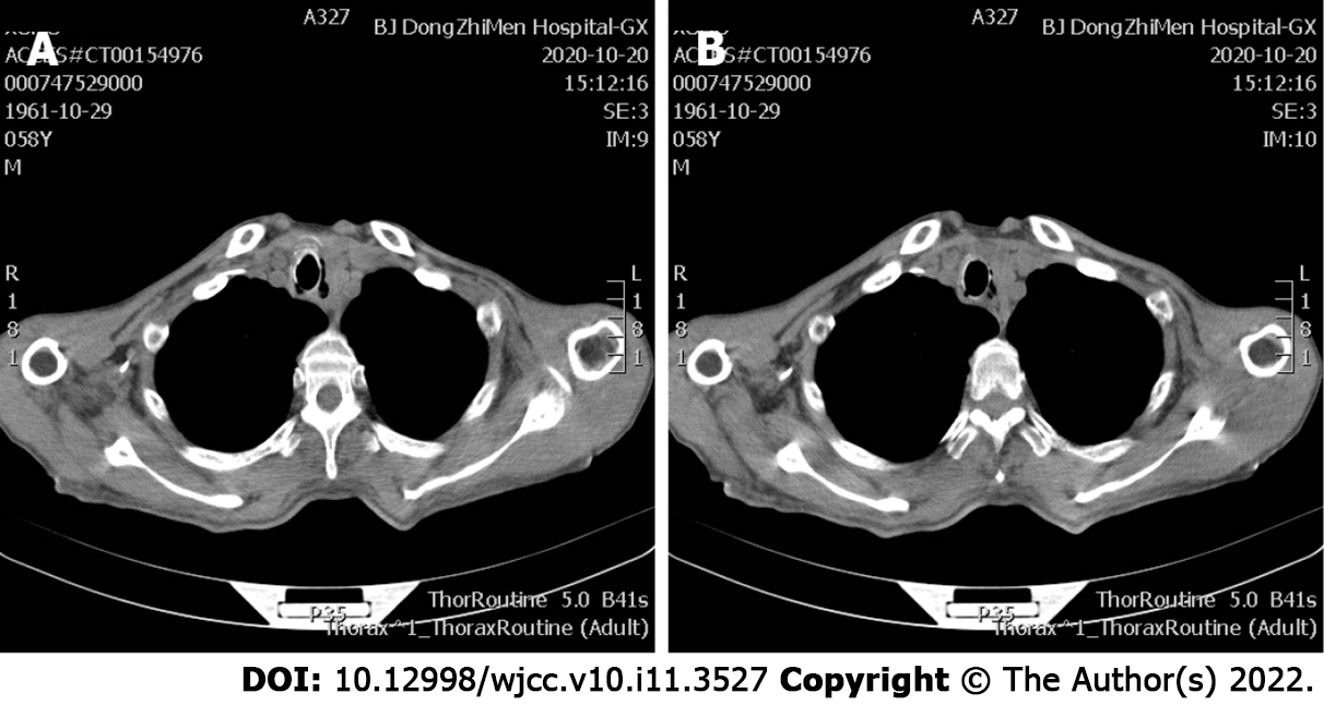
Figure 4 Computer tomography image showing the tracheoesophageal fistula after 4 mo of immunotherapy.
A: The progressed fistula after 4 mo of immunotherapy; B: A different slice image.
- Citation: Li CA, Yu WX, Wang LY, Zou H, Ban CJ, Wang HW. Double tracheal stents reduce side effects of progression of malignant tracheoesophageal fistula treated with immunotherapy: A case report. World J Clin Cases 2022; 10(11): 3527-3532
- URL: https://www.wjgnet.com/2307-8960/full/v10/i11/3527.htm
- DOI: https://dx.doi.org/10.12998/wjcc.v10.i11.3527


