Copyright
©The Author(s) 2022.
World J Clin Cases. Apr 16, 2022; 10(11): 3490-3495
Published online Apr 16, 2022. doi: 10.12998/wjcc.v10.i11.3490
Published online Apr 16, 2022. doi: 10.12998/wjcc.v10.i11.3490
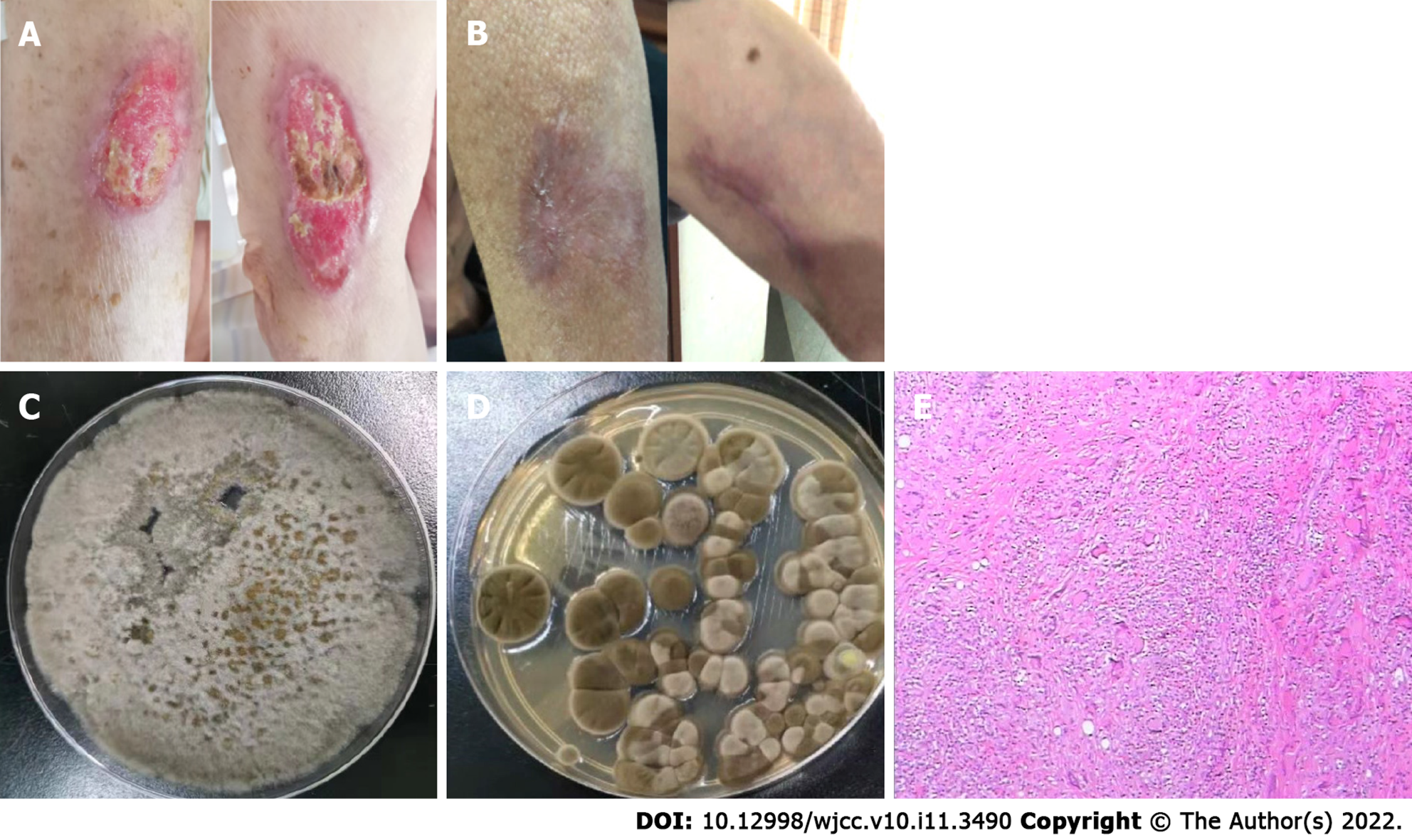
Figure 1 Symptoms, culture and pathology.
A: Hypertrophic erythema and deep ulcers on the left upper extremity; B: The ulcer on the left upper extremity had completely healed after treatment; C: Wound secretion culture revealed Corynespora cassiicola; D: Bronchoalveolar lavage fluid culture analysis revealed the presence of Cladosporium; E: Skin biopsy showed a partial squamous hyperplasia with a dermal granulomatous lesion.
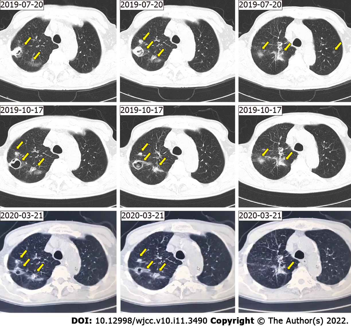
Figure 2 Imaging.
A chest computed tomography scan showing multiple nodules with multiple patchy areas in both lungs (arrow).
- Citation: Wang WY, Luo HB, Hu JQ, Hong HH. Pulmonary Cladosporium infection coexisting with subcutaneous Corynespora cassiicola infection in a patient: A case report. World J Clin Cases 2022; 10(11): 3490-3495
- URL: https://www.wjgnet.com/2307-8960/full/v10/i11/3490.htm
- DOI: https://dx.doi.org/10.12998/wjcc.v10.i11.3490


