Copyright
©The Author(s) 2022.
World J Clin Cases. Apr 16, 2022; 10(11): 3478-3484
Published online Apr 16, 2022. doi: 10.12998/wjcc.v10.i11.3478
Published online Apr 16, 2022. doi: 10.12998/wjcc.v10.i11.3478
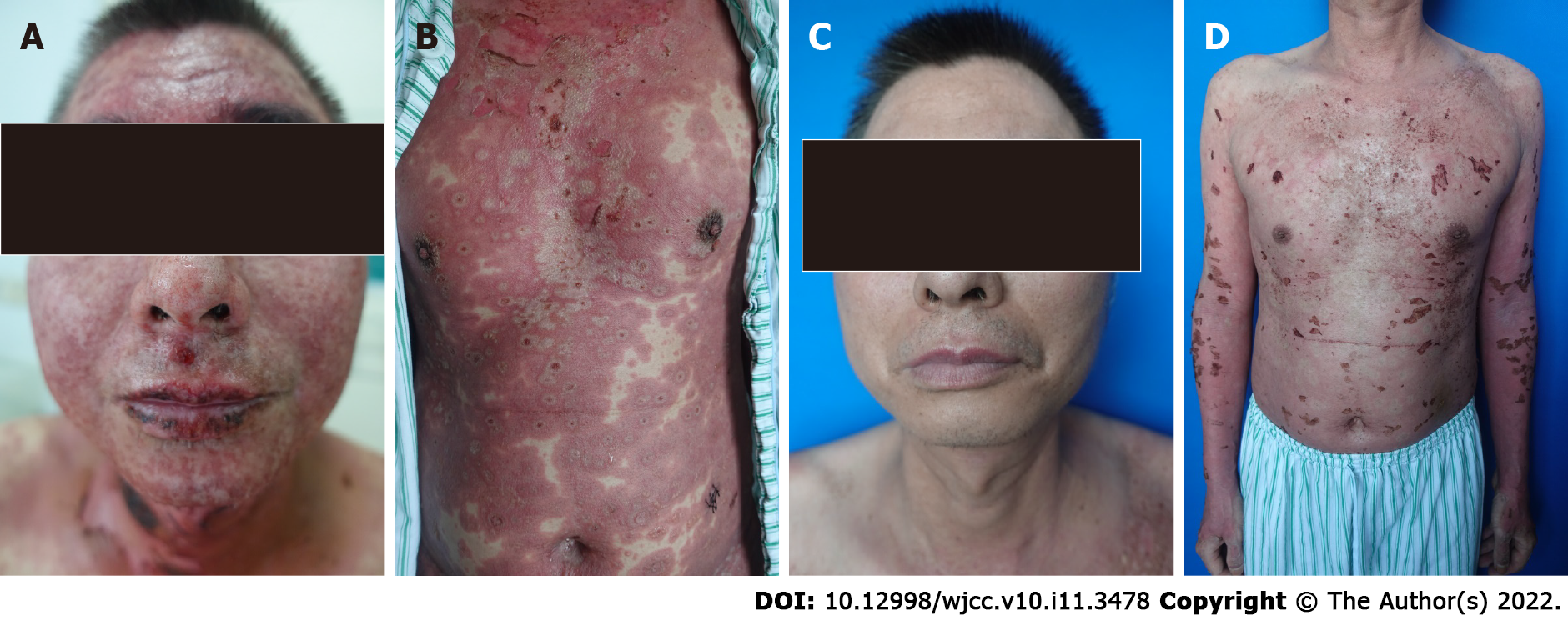
Figure 1 Clinical images before and after treatment.
A: Erythema, blisters and erosions appeared on the face and neck, as well as the oral mucosa; B: Typical erythema multiforme, slack bullae and epidermal peeling could be seen and covered more than 70% of the body surface area; C and D: The rashes gradually faded after 3 wk of treatment.
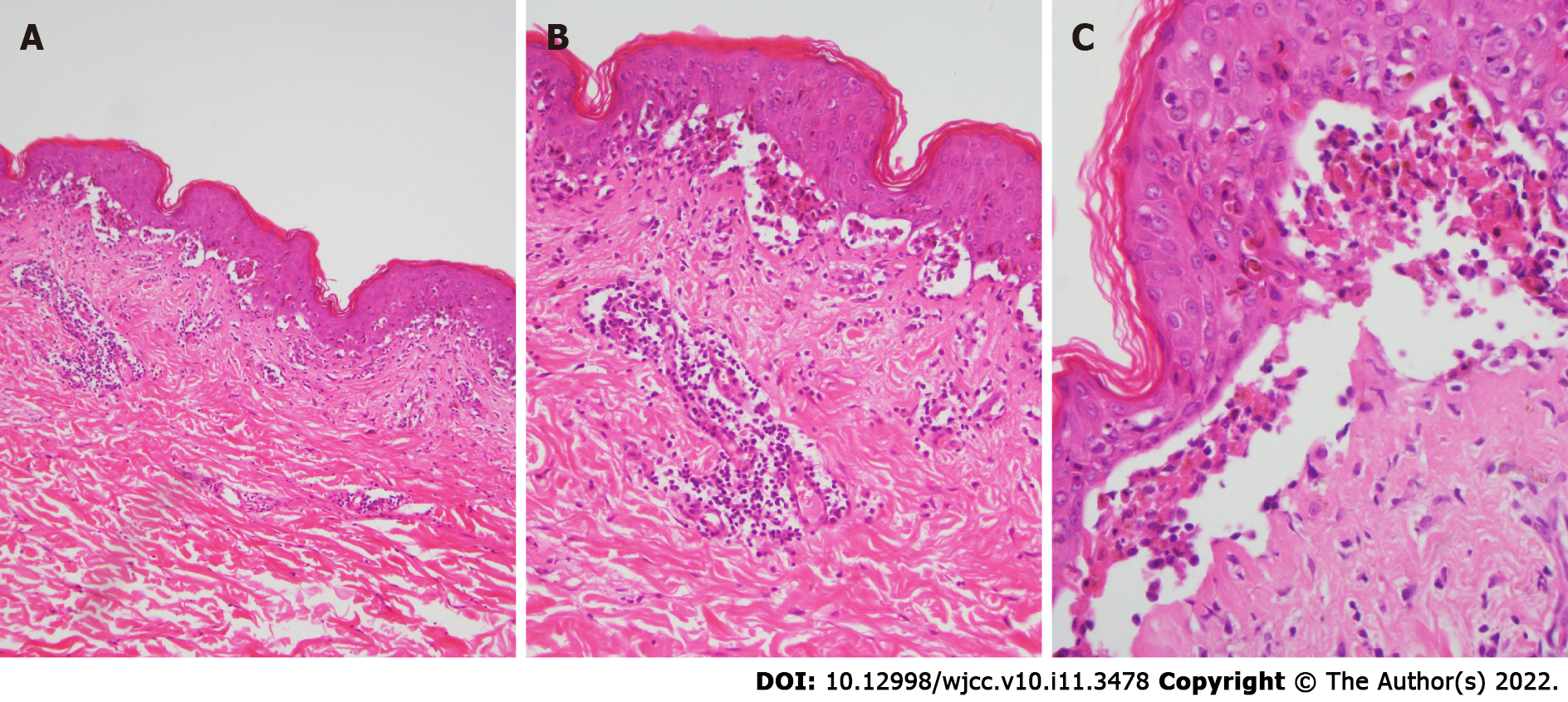
Figure 2 Pathological biopsy of the lesion.
A: Acantholytic bullae were observed in the epidermis [hematoxylin-eosin staining (HE) × 40]; B: Hyperkeratosis and liquefaction of basal cells could be seen in the epidermis. Blood vessels in the dermis were infiltrated by lymphocytes and eosinophils (HE × 100); C: Prominent necrotic keratinocytes were identified in the epidermis (HE × 400).
- Citation: Huang KK, Han SS, He LY, Yang LL, Liang BY, Zhen QY, Zhu ZB, Zhang CY, Li HY, Lin Y. Combination therapy (toripalimab and lenvatinib)-associated toxic epidermal necrolysis in a patient with metastatic liver cancer: A case report. World J Clin Cases 2022; 10(11): 3478-3484
- URL: https://www.wjgnet.com/2307-8960/full/v10/i11/3478.htm
- DOI: https://dx.doi.org/10.12998/wjcc.v10.i11.3478


