Copyright
©The Author(s) 2022.
World J Clin Cases. Apr 6, 2022; 10(10): 3297-3305
Published online Apr 6, 2022. doi: 10.12998/wjcc.v10.i10.3297
Published online Apr 6, 2022. doi: 10.12998/wjcc.v10.i10.3297
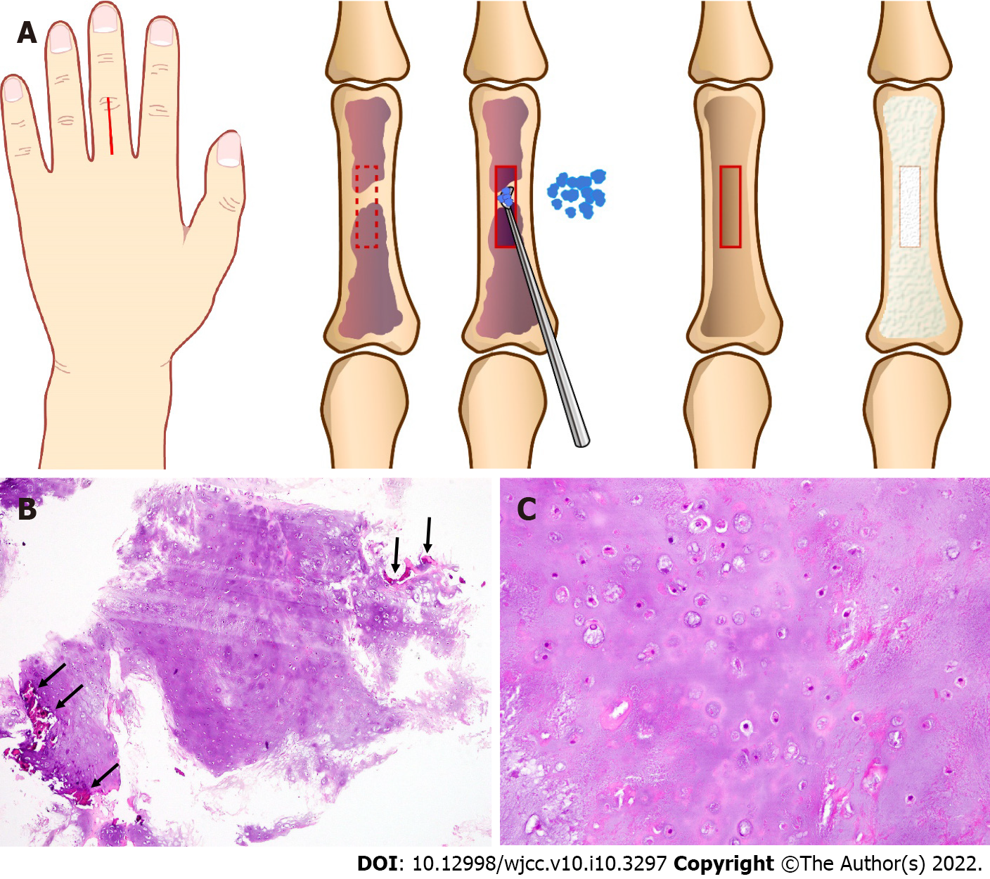
Figure 1 Schema and histological findings of the primary surgery at 76 years of age.
A: Schema of the primary surgery referencing the surgical record and figures. The tumor is located in the third proximal phalanx. After fenestration of the bone cortex, curettage and granular artificial bone grafting were performed under axillary nerve block. B: Magnification × 4. The tumor appears as a lobular, relatively cell-poor hyaline cartilage surrounded by an eosinophilic zone of reactive bone formation (encasement pattern; arrow). C: Magnification × 20. Scattered chondrocytes are located in sharp-edged lacunar spaces, with abundant hyaline cartilage matrix. Nuclei are small, round, and hyperchromatic, although larger, vesicular nuclei can also be present. No nuclear pleomorphism or enlargement is observed.
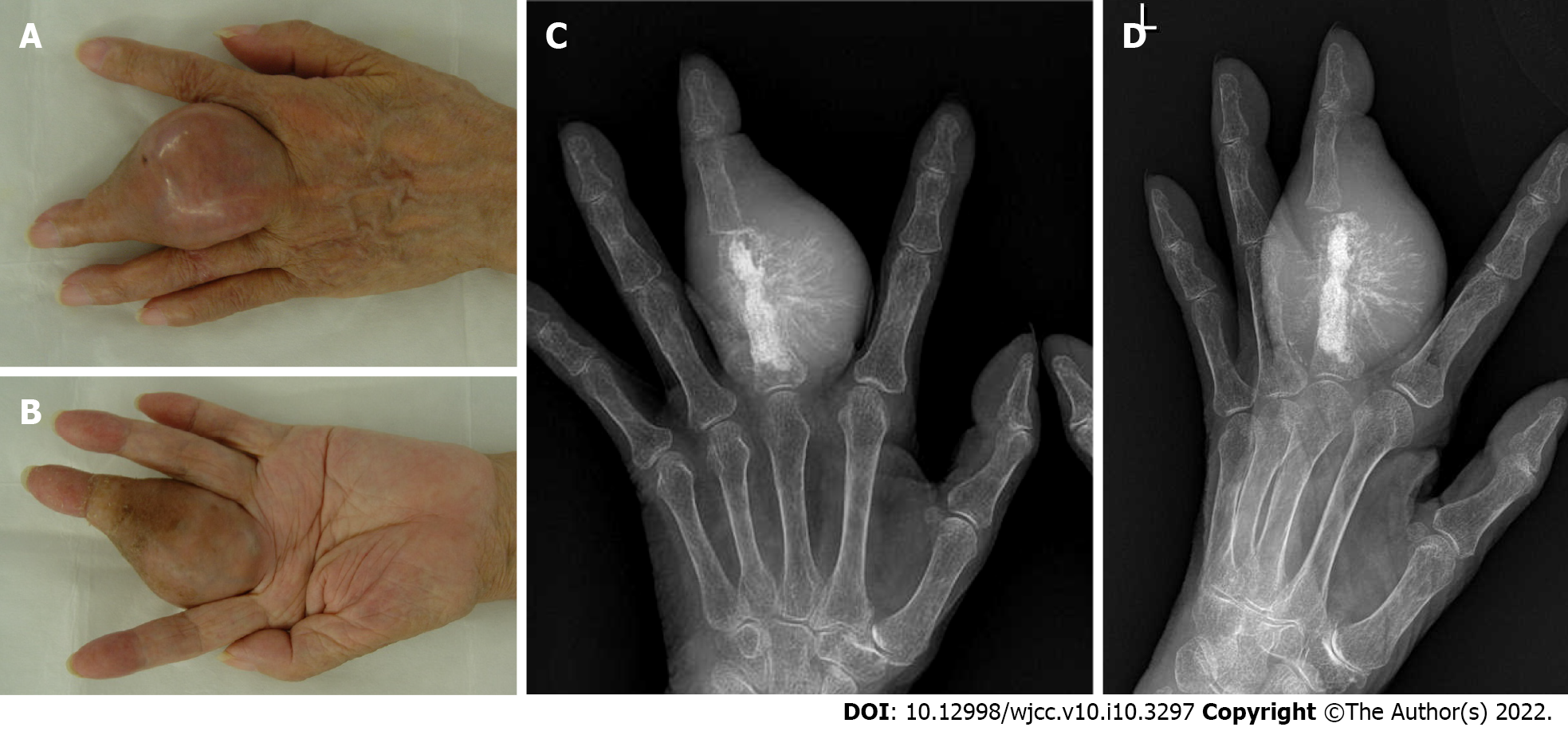
Figure 2 Clinical photographs and X-ray of the finger 11 years after the primary surgery.
A and B: Clinical photographs of the hand at 87 years of age. The left middle finger appears swollen and tense. The scar of the primary surgery is observed in the dorsal surface of the middle finger; A: Dorsal view; B: Palmar view; C and D: The X-ray shows residual artificial bone graft material and an expansive osteolytic lesion in the proximal phalanx of the middle finger; C: Anteroposterior view; D: Oblique view.
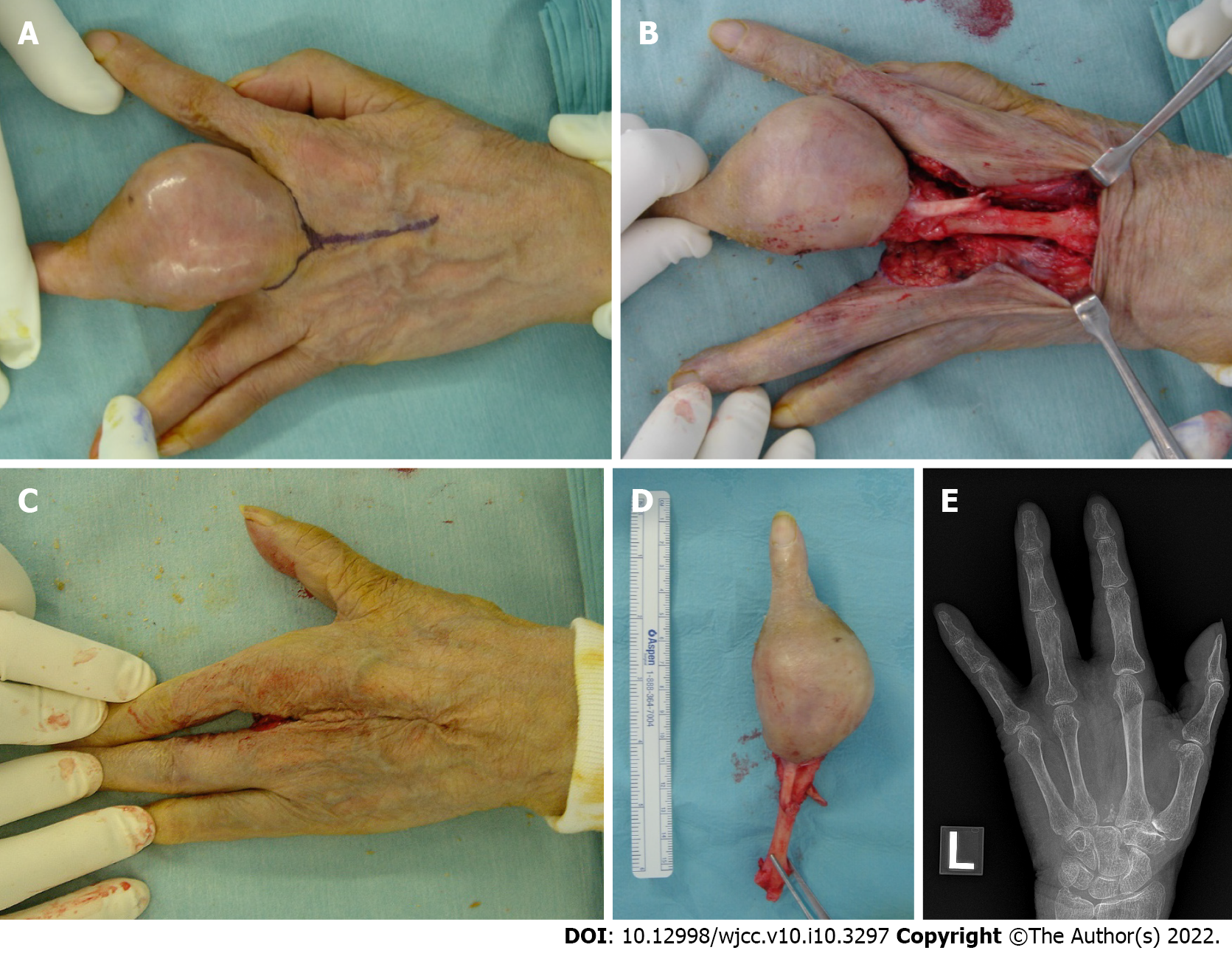
Figure 3 Intraoperative photographs and postoperative X-ray of the second surgery at 87 years of age.
A: A racket-shaped incision was planned; B: The third metacarpal bone is exposed and disarticulated at the carpometacarpal joint; C: The postoperative photograph of the dorsal aspect of the left hand demonstrates the gap closure; D: The amputated specimen; E: The postoperative X-ray of the left hand shows a good cosmetic four-finger hand.
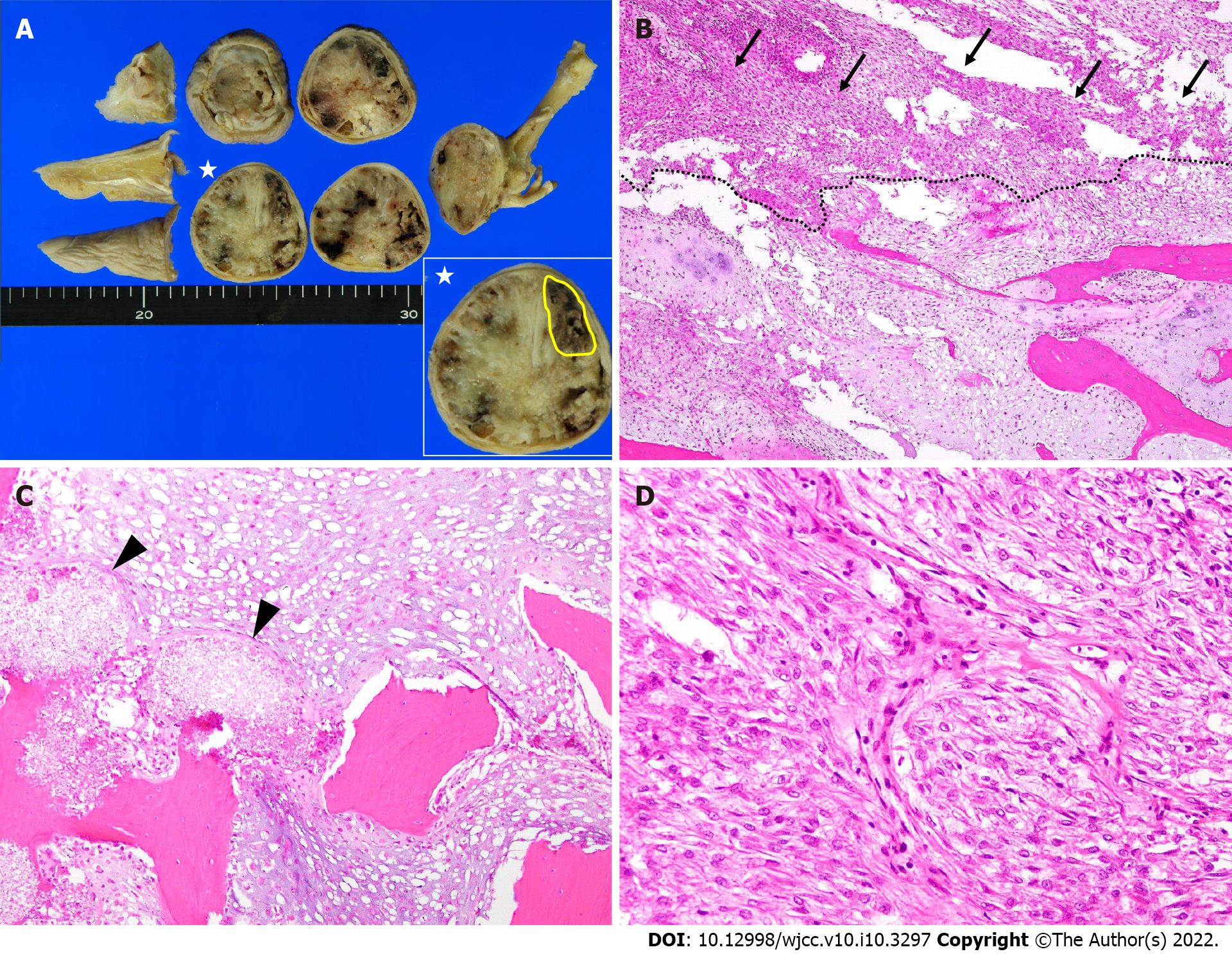
Figure 4 Histological findings of the amputated specimen.
A: Macroscopically, the tumor has destroyed the bone cortex and is extended to the soft tissue below the skin. The tumor is a grayish-white hyaline cartilage component filling the medullary cavity with hemorrhage and necrosis. In the largest cross-section of the tumor (star), the high-grade dedifferentiated component (yellow) accounts for 10.2% of the tumor; B: Magnification × 4. There is an abrupt transition between the low-grade chondrosarcoma (lower side) and high-grade dedifferentiated sarcoma components (upper side; arrow); C: Magnification × 10. The cartilaginous portion of the tumor shows a grade 1 or grade 2 chondrosarcoma with bone destruction. Arrowheads show the hydroxyapatite artificial bone graft that was used 11 years ago; D: Magnification × 20. The high-grade dedifferentiated component shows mitoses and atypia. The lesion is composed of spindle cells arranged in a fascicular growth pattern, resembling a high-grade undifferentiated pleomorphic sarcoma.
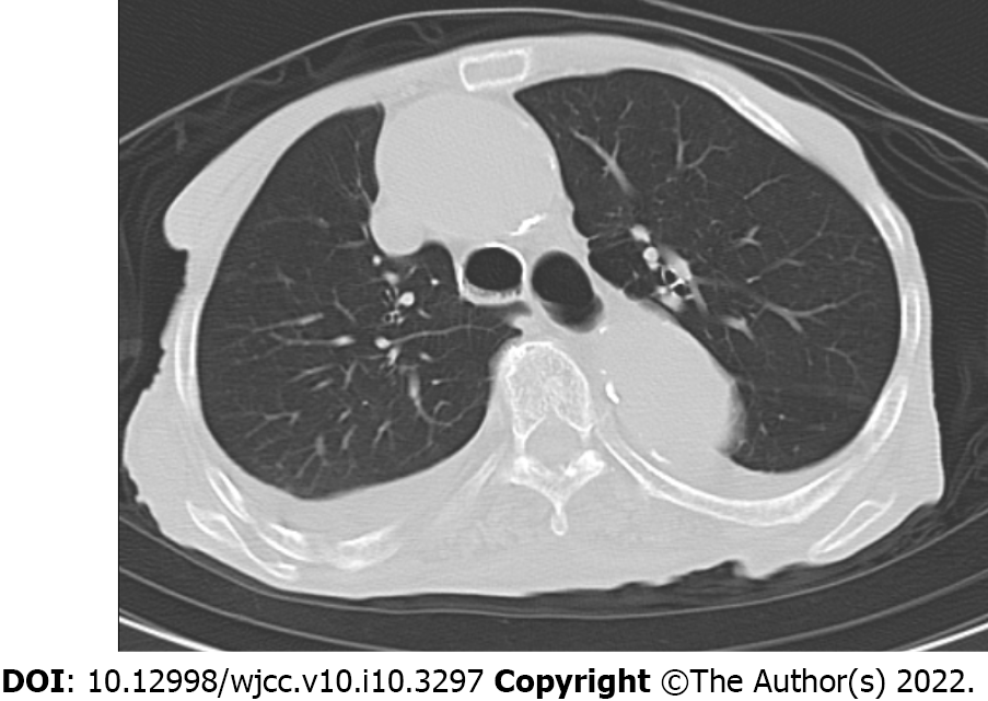
Figure 5 Computed tomogram of the 94-year-old woman, 6 years and 9 months after the second surgery.
The axial chest computed tomogram shows bilateral pleural effusion due to chronic congestive heart failure. No lung metastases are observed.
- Citation: Yonezawa H, Yamamoto N, Hayashi K, Takeuchi A, Miwa S, Igarashi K, Morinaga S, Asano Y, Saito S, Tome Y, Ikeda H, Nojima T, Tsuchiya H. Dedifferentiated chondrosarcoma of the middle finger arising from a solitary enchondroma: A case report. World J Clin Cases 2022; 10(10): 3297-3305
- URL: https://www.wjgnet.com/2307-8960/full/v10/i10/3297.htm
- DOI: https://dx.doi.org/10.12998/wjcc.v10.i10.3297


