Copyright
©The Author(s) 2022.
World J Clin Cases. Jan 7, 2022; 10(1): 79-90
Published online Jan 7, 2022. doi: 10.12998/wjcc.v10.i1.79
Published online Jan 7, 2022. doi: 10.12998/wjcc.v10.i1.79
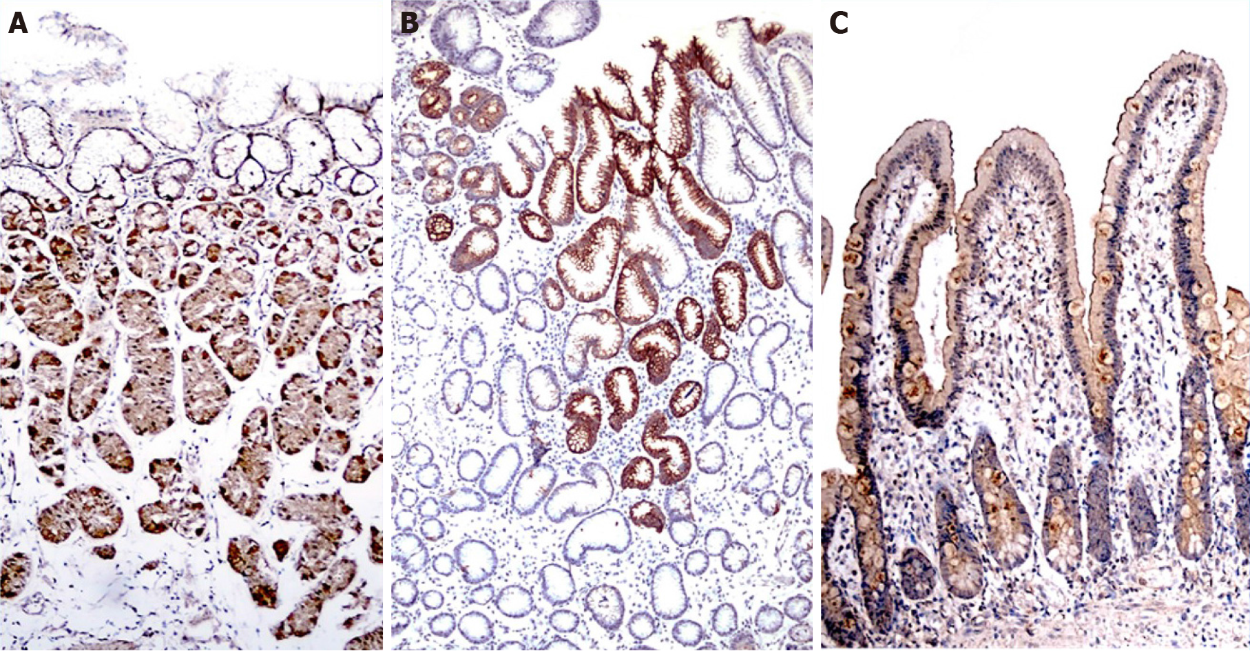
Figure 1 Distribution of transient receptor potential vanilloid-1 channels in the gastroduodenal mucosa were demonstrated by immunohistochemical staining.
A: Fundus; B: Antrum; C: Duodenum.
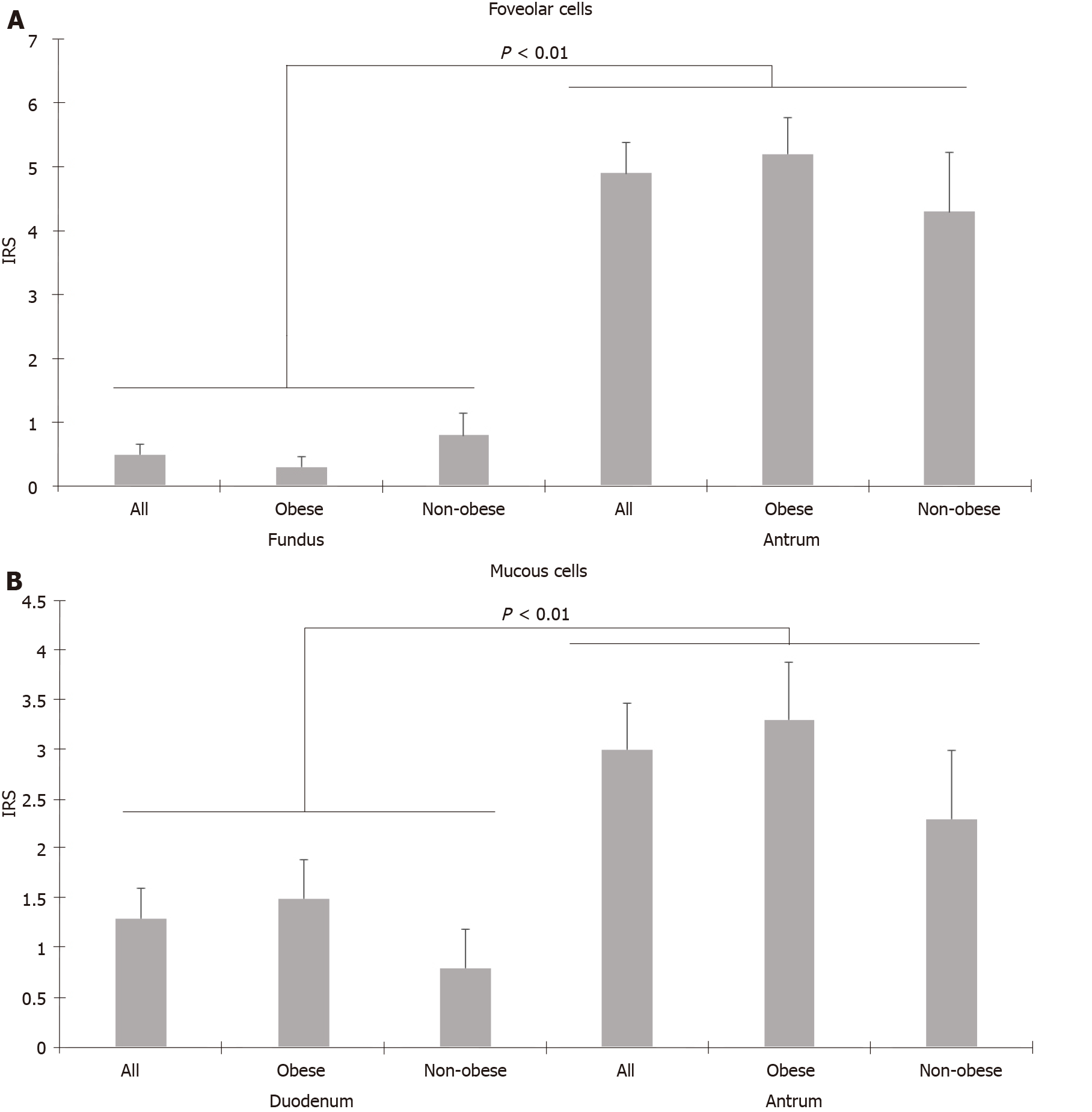
Figure 2 Comparison of immunoreactivity scores of foveolar cells in the fundus and antrum, mucous cells in the duodenum and antrum.
A: Foveolar cells in the fundus and antrum; B: Mucous cells in the duodenum and antrum. IRS: Immunoreactivity score.
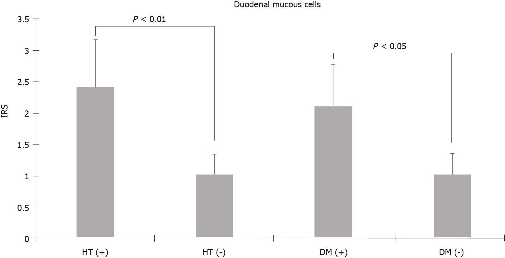
Figure 3 Duodenal mucous cells had higher immunoreactivity scores in patients with hypertension and diabetes.
IRS: Immunoreactivity score; HT (+): Hypertensive; HT (-): Normotensive; DM (+): Diabetic; DM (-): Non-diabetic.
- Citation: Atas U, Erin N, Tazegul G, Elpek GO, Yıldırım B. Distribution of transient receptor potential vanilloid-1 channels in gastrointestinal tract of patients with morbid obesity. World J Clin Cases 2022; 10(1): 79-90
- URL: https://www.wjgnet.com/2307-8960/full/v10/i1/79.htm
- DOI: https://dx.doi.org/10.12998/wjcc.v10.i1.79


