Copyright
©The Author(s) 2022.
World J Clin Cases. Jan 7, 2022; 10(1): 361-370
Published online Jan 7, 2022. doi: 10.12998/wjcc.v10.i1.361
Published online Jan 7, 2022. doi: 10.12998/wjcc.v10.i1.361
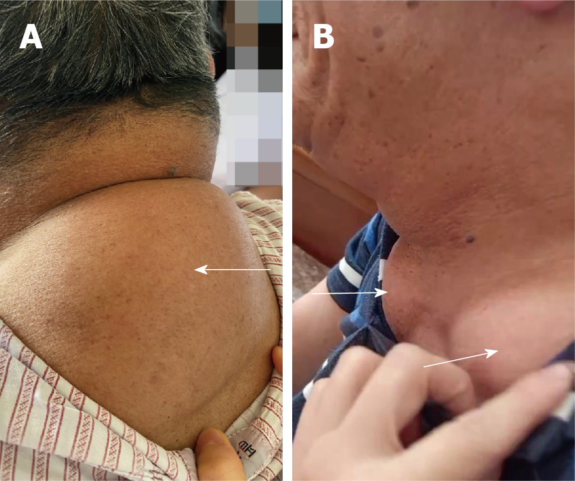
Figure 1 Masses in the patient's upper back and neck.
A: Masses in the upper back (white arrow); B: Masses in each supraclavicular fossa (white arrows).
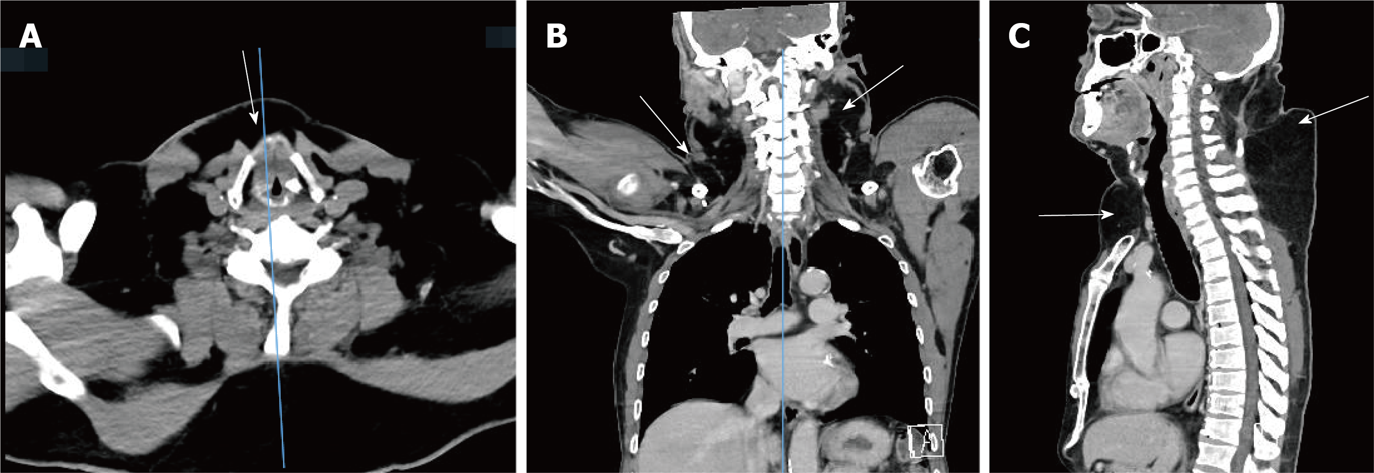
Figure 2 Contrast-enhanced computed tomographic images of the patient’s neck and chest.
A: The axial view revealed anterior neck masses (white arrow); B: The coronal view showed lateral neck masses (white arrows); C: The sagittal view showed masses in the anterior neck, sternum and supraclavicular fossa and upper back (white arrows).
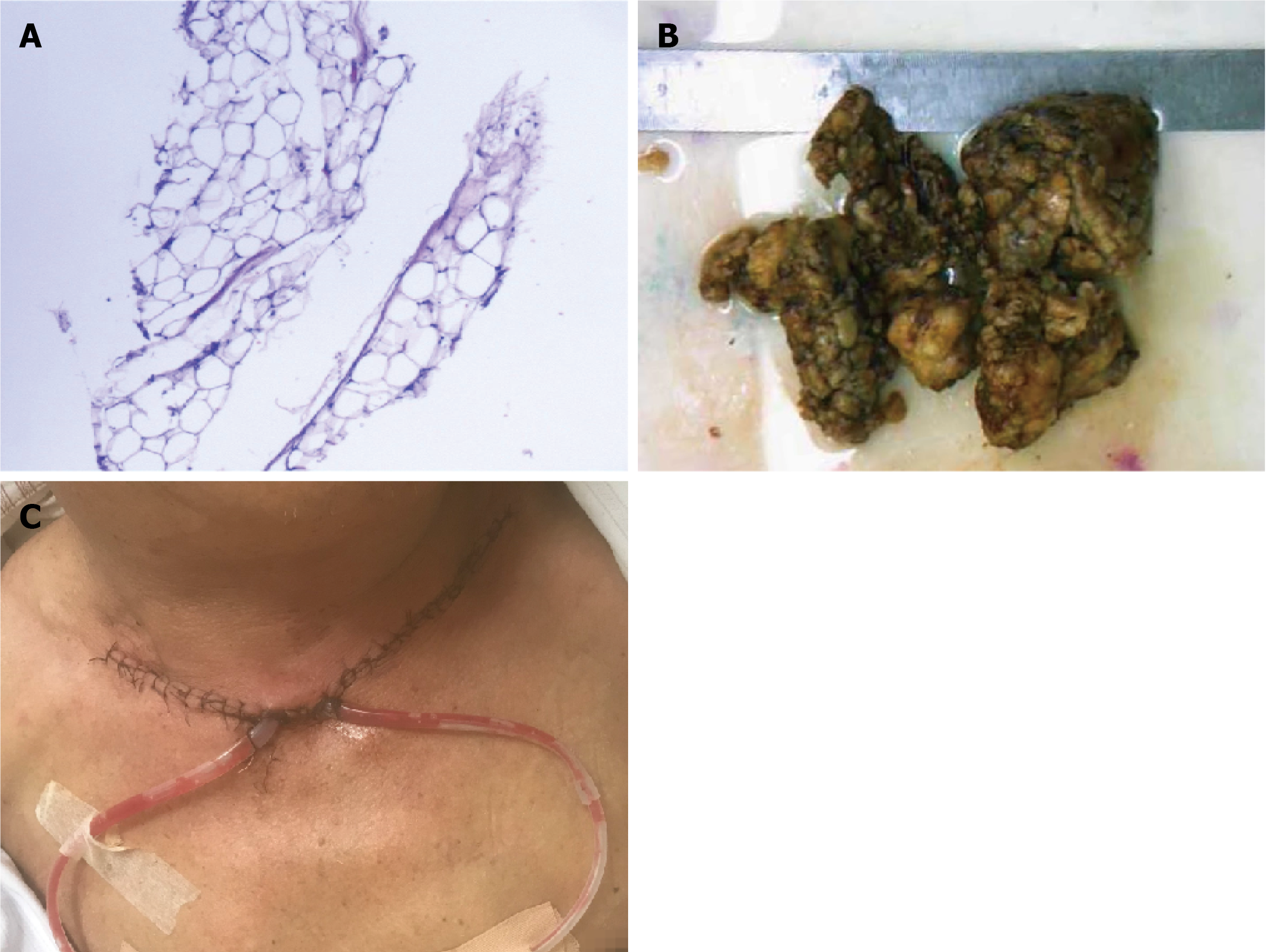
Figure 3 Histopathologic images and postoperative clinical appearance of the masses.
A: Histopathologic image (200 ×); B: Resected mass image; C: Postoperative area image.
- Citation: Yan YJ, Zhou SQ, Li CQ, Ruan Y. Diagnostic and surgical challenges of progressive neck and upper back painless masses in Madelung’s disease: A case report and review of literature. World J Clin Cases 2022; 10(1): 361-370
- URL: https://www.wjgnet.com/2307-8960/full/v10/i1/361.htm
- DOI: https://dx.doi.org/10.12998/wjcc.v10.i1.361


