Copyright
©The Author(s) 2022.
World J Clin Cases. Jan 7, 2022; 10(1): 345-352
Published online Jan 7, 2022. doi: 10.12998/wjcc.v10.i1.345
Published online Jan 7, 2022. doi: 10.12998/wjcc.v10.i1.345
Figure 1 Multiple nodules over the left breast and chest wall.
A: Nodules measuring 1-2 cm in size; B: Status of the skin lesions at 3 mo after endocrine therapy with 25 mg of exemestane and 10 mg of everolimus; C: After 8 mo of endocrine therapy, the metastatic skin lesions progressed.
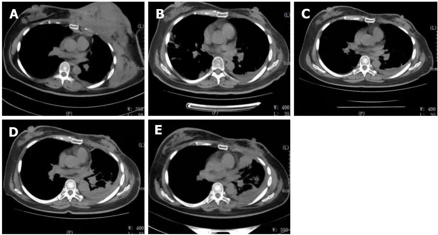
Figure 2 Changes in thoracic computed tomography manifestations in the patient throughout the treatment period.
A: Thoracic computed tomography showed a mass in the left breast and chest wall muscles before treatment; B: Partial response was clinically observed after two cycles of chemotherapy. However, the pleural effusion increased; C: Breast and chest wall tumors showed a significant response after 1 mo of endocrine therapy; D: The breast and chest wall tumors progressed, and the pleural effusion increased again; E: After two cycles of chemotherapy, the efficacy was evaluated as stable.
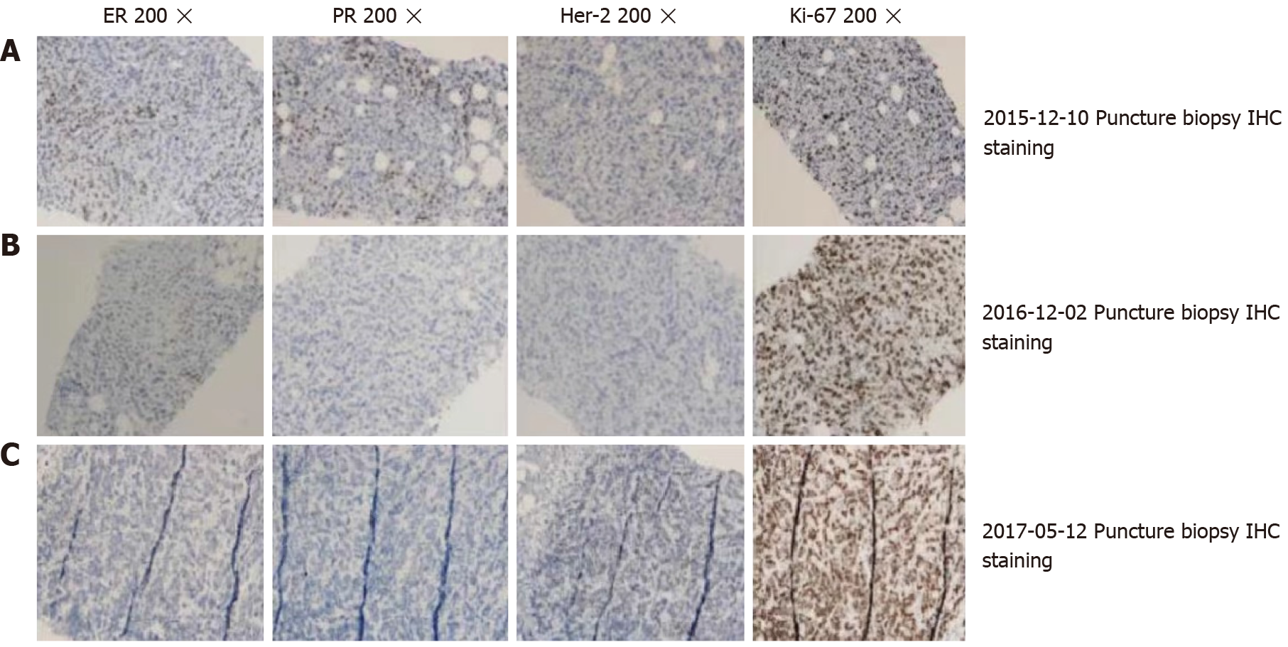
Figure 3 Immunohistochemical results at different puncture time points.
A: Immunohistochemical staining of the breast puncture biopsy (20 × magnification); B: High expression of ER (+), PR (+), and Ki-67 (40%), and low expression of Her-2 (-); C: Triple negative expression of ER (-), PR (-), and HER2 (-), and high expression of Ki-67 (60%); D: Low expression of ER (10%+), PR (-), HER2 (-), and high expression of Ki-67 (90%). The pathology results in A, B, and C were obtained from the same patient at different time points of puncture.
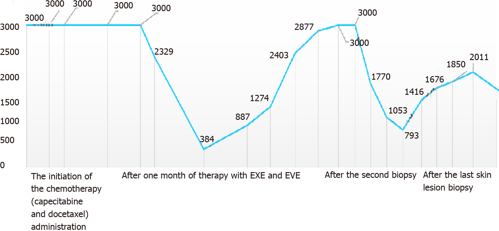
Figure 4 Changes of the tumor marker CA15-3 in the treatment process.
The initiation of the chemotherapy (capecitabine and docetaxel) administration. The CA15-3 serum concentration continued to be above the upper limit of the reference value. After one month of therapy with EXE and EVE, the CA15-3 serum concentration immediately and sharply declined. After the second biopsy, the CA15-3 serum concentration decreased when adriamycin plus cyclophosphamide chemotherapy was administered. The trend for the tumor markers in the other treatments after the last skin lesion biopsy is shown. EXE: Exemestane; EVE: Everolimus.
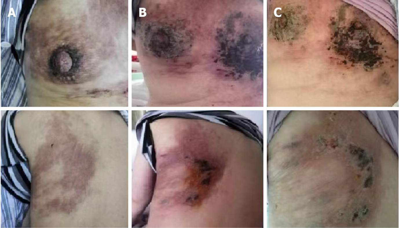
Figure 5 Skin changes before, during, and after the patient received photodynamic therapy (PDT) and sonodynamic therapy (SDT).
A: Before performing photodynamic therapy (PDT) followed by sonodynamic therapy (SDT); B: At 3 d after PDT and SDT treatment for the skin lesion, the skin lesion presented with necrosis; C: The lesions had rapid improvement with necrosis, and crusting after 10 d of PDT and SDT treatment.
- Citation: Li ZH, Wang F, Zhang P, Xue P, Zhu SJ. Diagnosis and guidance of treatment of breast cancer cutaneous metastases by multiple needle biopsy: A case report. World J Clin Cases 2022; 10(1): 345-352
- URL: https://www.wjgnet.com/2307-8960/full/v10/i1/345.htm
- DOI: https://dx.doi.org/10.12998/wjcc.v10.i1.345


