Copyright
©The Author(s) 2022.
World J Clin Cases. Jan 7, 2022; 10(1): 155-165
Published online Jan 7, 2022. doi: 10.12998/wjcc.v10.i1.155
Published online Jan 7, 2022. doi: 10.12998/wjcc.v10.i1.155
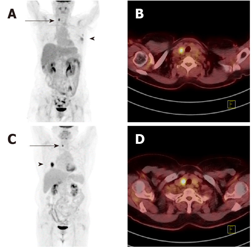
Figure 1 Examples of focal hypermetabolic thyroid incidentaloma.
A: Focal fluorodeoxyglucose (FDG) uptake is observed in the right lower neck on the maximum intensity projection (MIP) image of a 53-year-old woman diagnosed with left breast cancer (FDG uptake in the left breast and axillary fossa); B: On the axial view, the focal FDG uptake is observed in the right thyroid lobe with maximum standardized uptake value (SUVmax) 5.9 and it was diagnosed as a benign nodule by cytological/histological examination; C: Focal FDG uptake is observed in the right lower neck on the MIP image of a 66-year-old woman diagnosed with adenocarcinoma in the right lower lobe of lung as a result of biopsy performed due to abnormal radiologic findings; D: On the axial view, the focal FDG uptake is observed in the right thyroid lobe (SUVmax 8.6) and the cytological/histological examination revealed papillary thyroid cancer.
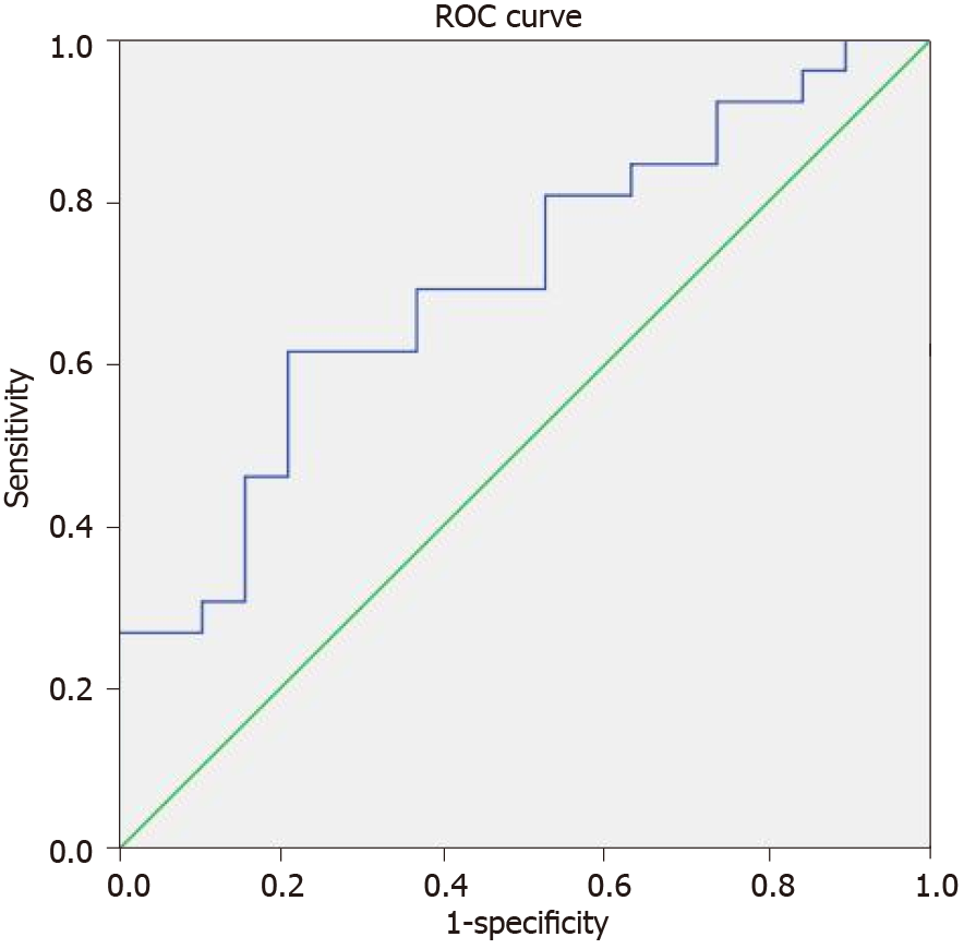
Figure 2 The receiver operating characteristic curve for maximum standardized uptake value.
The area under the curve is 0.702 (95% confidence interval, lower: 0.550, upper: 0.855). The cut-off value for maximum standardized uptake value is 8.5 with a sensitivity of 0.615 and a specificity of 0.789. ROC: Receiver operating characteristic.
- Citation: Lee H, Chung YS, Lee JH, Lee KY, Hwang KH. Characterization of focal hypermetabolic thyroid incidentaloma: An analysis with F-18 fluorodeoxyglucose positron emission tomography/computed tomography parameters. World J Clin Cases 2022; 10(1): 155-165
- URL: https://www.wjgnet.com/2307-8960/full/v10/i1/155.htm
- DOI: https://dx.doi.org/10.12998/wjcc.v10.i1.155


