Copyright
©2013 Baishideng Publishing Group Co.
World J Clin Cases. Jul 16, 2013; 1(4): 146-148
Published online Jul 16, 2013. doi: 10.12998/wjcc.v1.i4.146
Published online Jul 16, 2013. doi: 10.12998/wjcc.v1.i4.146
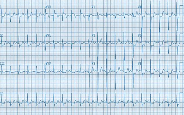
Figure 1 Electrocardiogram shows sinus tachycardia with a rate of 130, 1 mm to 3 mm ST depressions in leads I, II, and V3 to V6 and 1 mm to 2 mm ST elevations in aVR and V1.
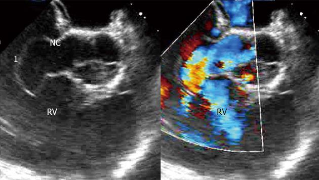
Figure 2 Subsequent transesophageal echocardiogram done, showed a ruptured aneurysm of the noncoronary sinus of Valsalva, that is Windsock in morphology, into the right atrium.
A: Sinus of Valsalva defect with; B: Color-Doppler. 1: Windsock aneurysm; RV: Right ventricle; NC: Noncoronary cusp.
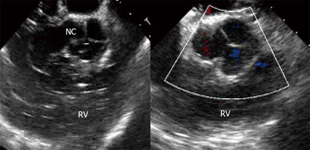
Figure 3 Patient underwent immediately surgery to repair both the ruptured aneurysm of the noncoronary sinus of Valsalva and the ventricular septal defect.
A: Bubbles going through membranous ventricular septal defect; B: The repaired defect. Asterisk indicates micro air bubble traversing the membranous VSD. RV: Right ventricle; NC: Noncoronary cusp.
- Citation: Cao LB, Hannon D, Movahed A. Noncoronary sinus of Valsalva rupture into the right atrium with a coexisting perimembranous ventricular septal defect. World J Clin Cases 2013; 1(4): 146-148
- URL: https://www.wjgnet.com/2307-8960/full/v1/i4/146.htm
- DOI: https://dx.doi.org/10.12998/wjcc.v1.i4.146


