Copyright
©2013 Baishideng Publishing Group Co.
World J Clin Cases. Jun 16, 2013; 1(3): 128-133
Published online Jun 16, 2013. doi: 10.12998/wjcc.v1.i3.128
Published online Jun 16, 2013. doi: 10.12998/wjcc.v1.i3.128
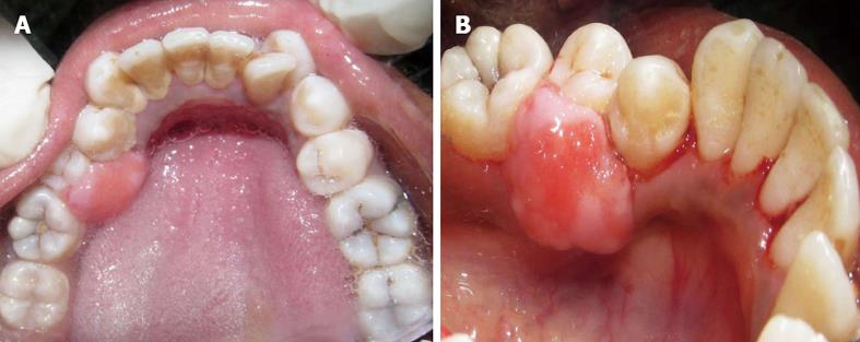
Figure 1 Lingual view of the lesion with smooth non-ulcerated surface and broad attachment base.
A: The lesion with smooth non-ulcerated surface; B: Broad attachment base.
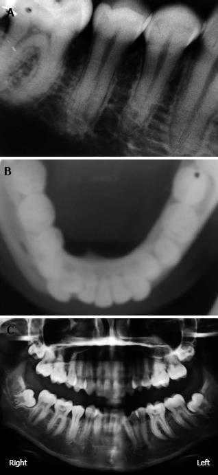
Figure 2 Non involvement of bone.
A: Intra oral periapical radiograph; B: Maxillary occlusal radiograph; C: Ortho pantomo radiograph.
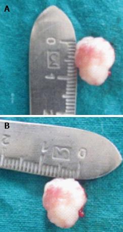
Figure 3 Excised tissue measuring 1.
5 cm × 1 cm. A: Length 1.5 cm; B: Width 1 cm.
Figure 4 Interrupted sutures placed with 3-0 black braided silk.
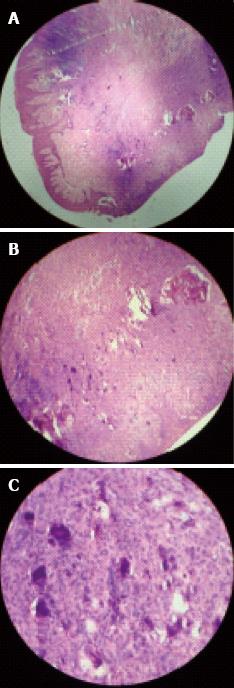
Figure 5 The features were suggestive of “peripheral cemento-ossifying fibroma”.
A: Histological picture showing long slender rete ridges and parakeratinized stratified squamous epithelium; B: Round to ovoid basophilic cementum-like calcifications [hematoxylin-eosin (HE) staining, × 10]; C: Basophilic globules of calcified mass along with osteoid tissue, round to ovoid basophilic cementum-like calcifications (HE staining, × 40).
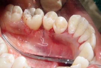
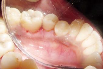
Figure 6 Post operative photograph of surgical site showing satisfactory healing 30 d after surgery.
- Citation: Mishra AK, Maru R, Dhodapkar SV, Jaiswal G, Kumar R, Punjabi H. Peripheral cemento-ossifying fibroma: A case report with review of literature. World J Clin Cases 2013; 1(3): 128-133
- URL: https://www.wjgnet.com/2307-8960/full/v1/i3/128.htm
- DOI: https://dx.doi.org/10.12998/wjcc.v1.i3.128


