Copyright
©2013 Baishideng.
World J Clin Cases. Apr 16, 2013; 1(1): 49-51
Published online Apr 16, 2013. doi: 10.12998/wjcc.v1.i1.49
Published online Apr 16, 2013. doi: 10.12998/wjcc.v1.i1.49
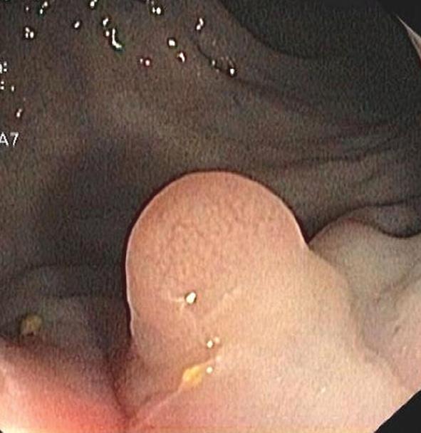
Figure 1 In case of a rectal schwannoma, the colonoscopy disclosed evidence of a little polypoid lesion in the left lateral wall of the rectum.
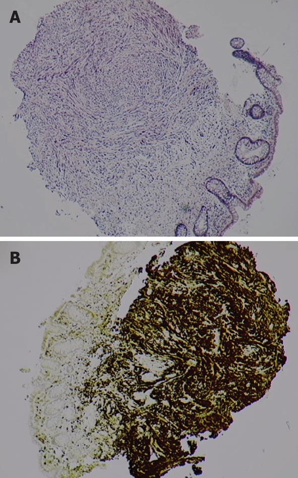
Figure 2 In case of rectal schwannoma, the tumor, located in the submucosa, has a nodular well-defined structure (haematoxylin and eosin, ×20).
The structure that consists in proliferation of Schwann-like cells, showing a palisading arrangement (A), being marked by S-100 protein (B).
- Citation: Zippi M, Pica R, Scialpi R, Cassieri C, Avallone EV, Occhigrossi G. Schwannoma of the rectum: A case report and literature review. World J Clin Cases 2013; 1(1): 49-51
- URL: https://www.wjgnet.com/2307-8960/full/v1/i1/49.htm
- DOI: https://dx.doi.org/10.12998/wjcc.v1.i1.49


