Copyright
©The Author(s) 2024.
World J Clin Oncol. Aug 24, 2024; 15(8): 1110-1116
Published online Aug 24, 2024. doi: 10.5306/wjco.v15.i8.1110
Published online Aug 24, 2024. doi: 10.5306/wjco.v15.i8.1110
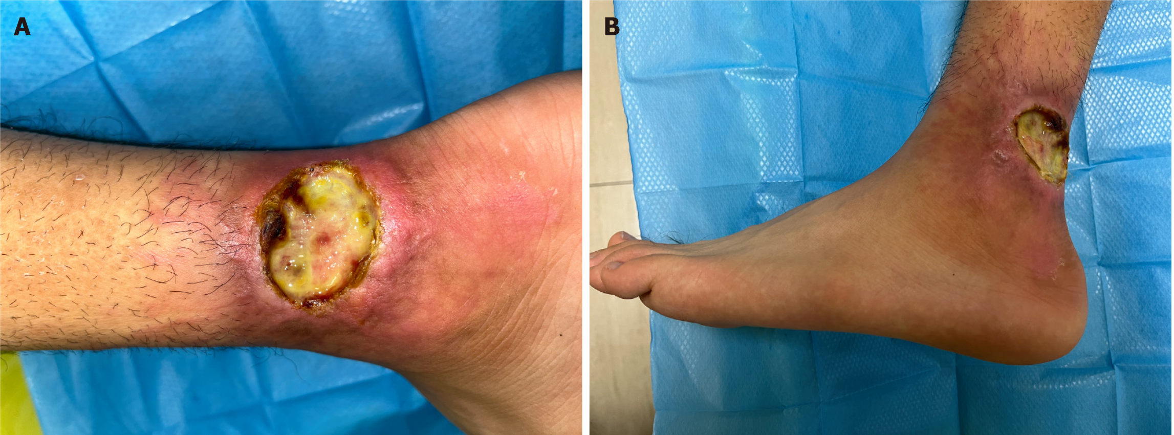
Figure 1 Poorly healed wound caused by non-Hodgkin's lymphoma from the patient's skin.
A: Localized wound on the left calf near the ankle; B: Overall view of the wound located on the left calf near the ankle. A persistent skin ulcer with purulent discharge near the left lower extremity ankle and redness and swelling of the surrounding skin.
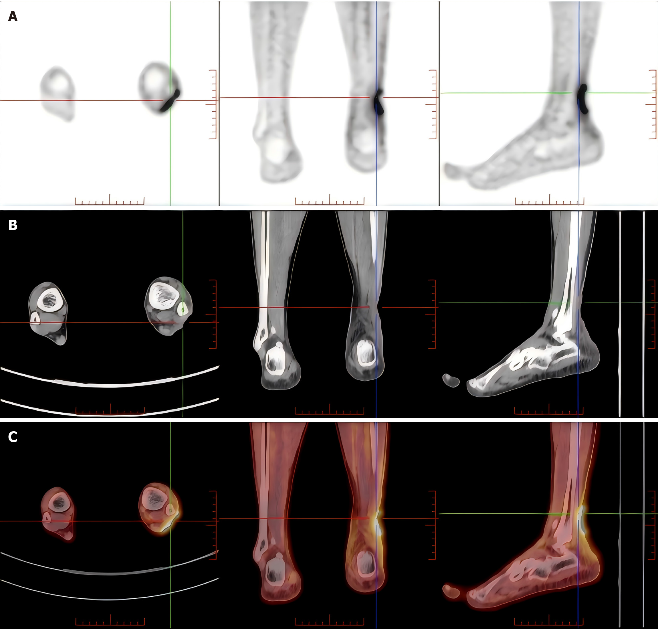
Figure 2 Whole-body positron emission tomography/computed tomography imaging with 18F-fluorodeoxyglucose before surgical treatment.
A: Positron emission tomography (PET) images of the calf in cross-sectional, coronal, and sagittal views; B: computed tomography (CT) images of the calf in cross-sectional, coronal, and sagittal views; C: PET-CT fusion images of the calf in cross-sectional, coronal, and sagittal views. Significant uptake of 18F-fluorodeoxyglucose in the left distal calf near the outer ankle wound, indicating increased metabolic activity.
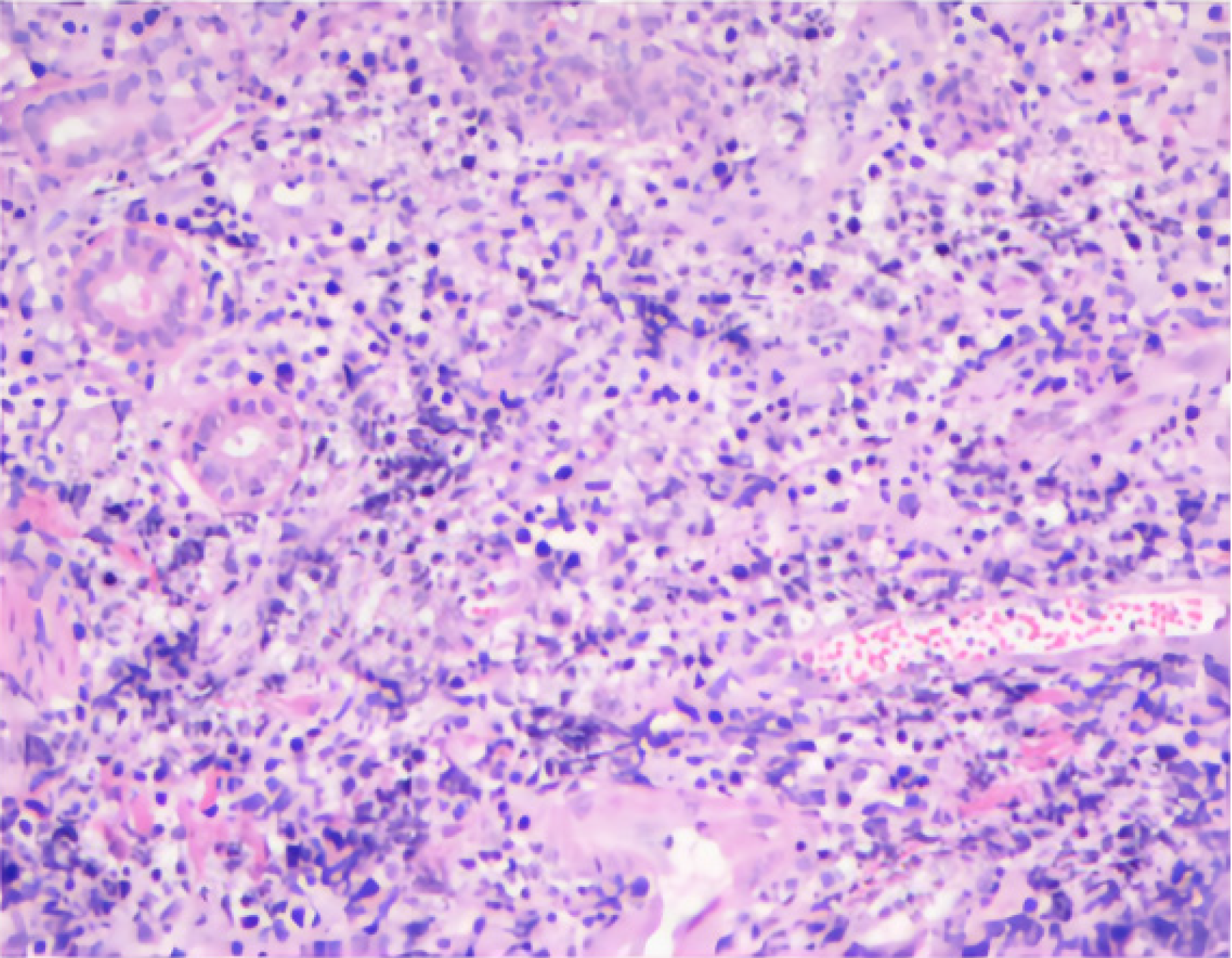
Figure 3 Histopathologic analysis of preoperative wound specimens.
Some of the epithelium undergoes exfoliation and necrosis, and heterogeneous cells are observed in the dermis, infiltrating the skin in a multifocal manner, predominantly around blood vessels and skin appendages.
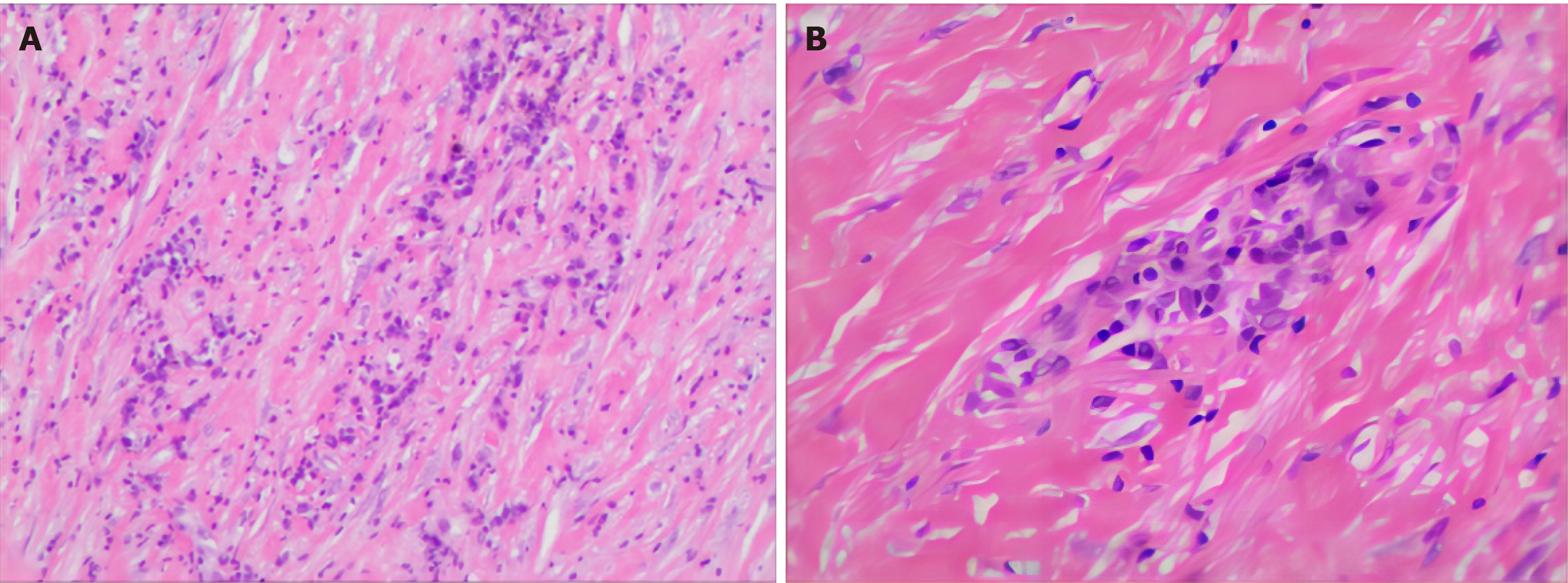
Figure 4 Hematoxylin and eosin staining of postoperative margin tissue specimens.
A: low magnification; B: High magnification. A large plasma cell and lymphocyte infiltrate was seen around the vessels, consistent with a chronic ulcerative lesion with marked vessel wall thickening and lumen occlusion, with no evidence of a neoplastic lesion.
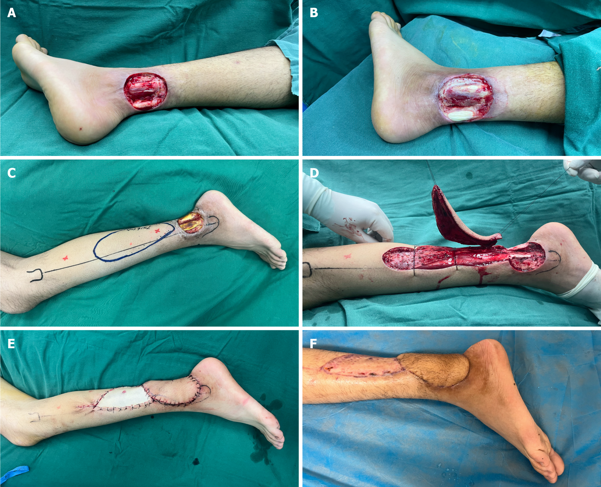
Figure 5 Surgical outcomes of chronic difficult-to-heal wounds in the patient with non-Hodgkin's lymphoma.
A: Appearance of wounds after extended excision debridement; B: Appearance of wounds after multiple debridements; C: Preoperative appearance of wounds and design of the flap; D: Flap cutting; E: Appearance of flap suturing after surgery; F: 1-month follow-up after flap surgery, with good texture of the flap, and no rupture or infection.
Figure 6 Skin changes after 1 year of wound treatment for non-Hodgkin's lymphoma.
A: The inner side of the left calf; B: The lateral side of the left calf. The wound flap healed well and no lymphoma recurrence was seen.
- Citation: Zhang PS, Wang R, Wu HW, Zhou H, Deng HB, Fan WX, Li JC, Cheng SW. Non-Hodgkin's lymphoma involving chronic difficult-to-heal wounds: A case report. World J Clin Oncol 2024; 15(8): 1110-1116
- URL: https://www.wjgnet.com/2218-4333/full/v15/i8/1110.htm
- DOI: https://dx.doi.org/10.5306/wjco.v15.i8.1110


