Copyright
©The Author(s) 2022.
World J Cardiol. Feb 26, 2022; 14(2): 64-82
Published online Feb 26, 2022. doi: 10.4330/wjc.v14.i2.64
Published online Feb 26, 2022. doi: 10.4330/wjc.v14.i2.64
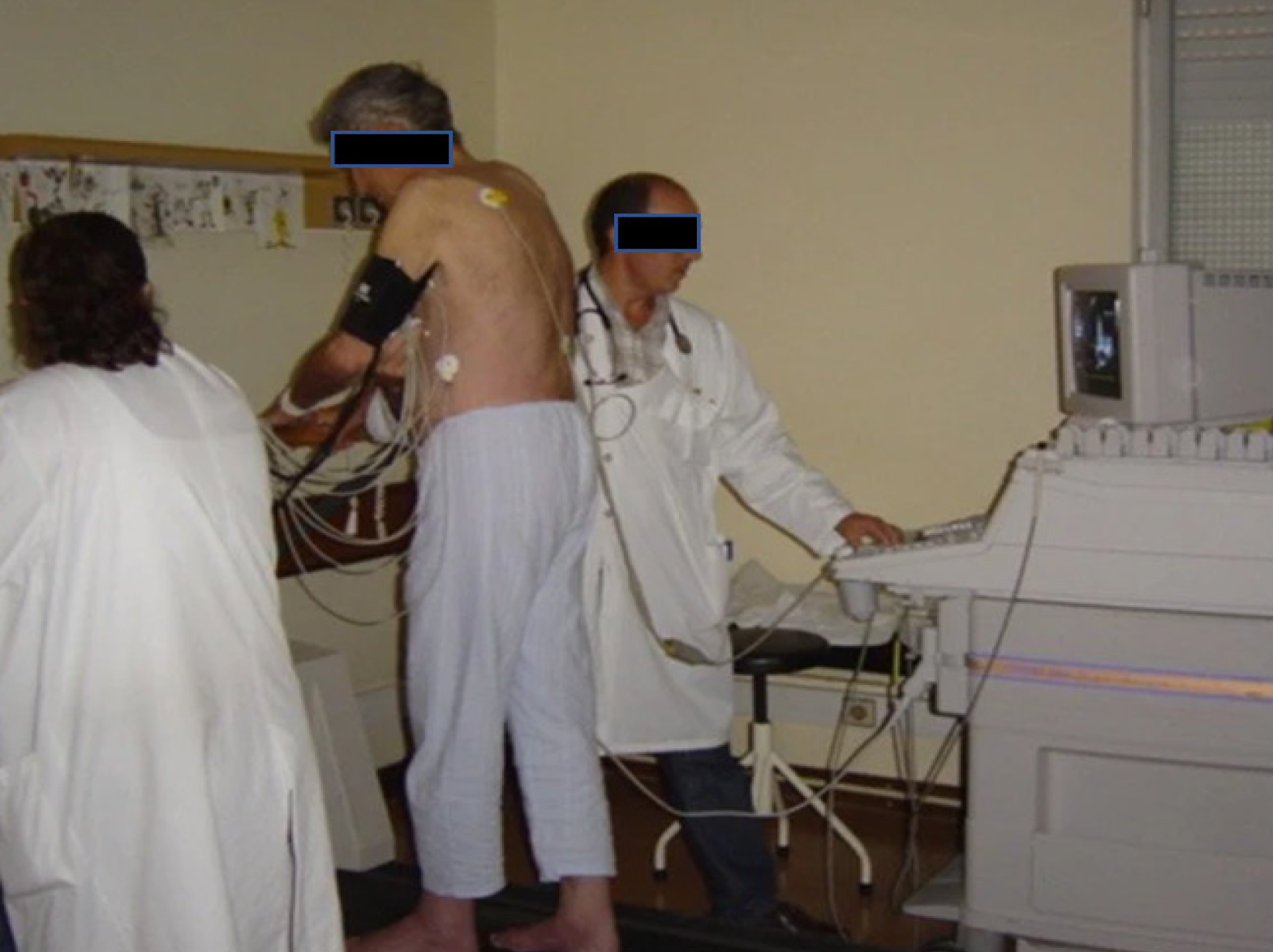
Figure 1 Echocardiographic data acquisition with the patient in the orthostatic position during exercise on a treadmil.
Citation: Cotrim C, João I, Fazendas P, Almeida AR, Lopes L, Stuart B, Cruz I, Caldeira D, Loureiro MJ, Morgado G, Pereira H. Clinical applications of exercise stress echocardiography in the treadmill with upright evaluation during and after exercise. Cardiovasc Ultrasound 2013; 11: 26 [PMID: 23875614 DOI: 10.1186/1476-7120-11-26] Copyright © The Author (s) 2013. Published by BMC part of Springer Nature.
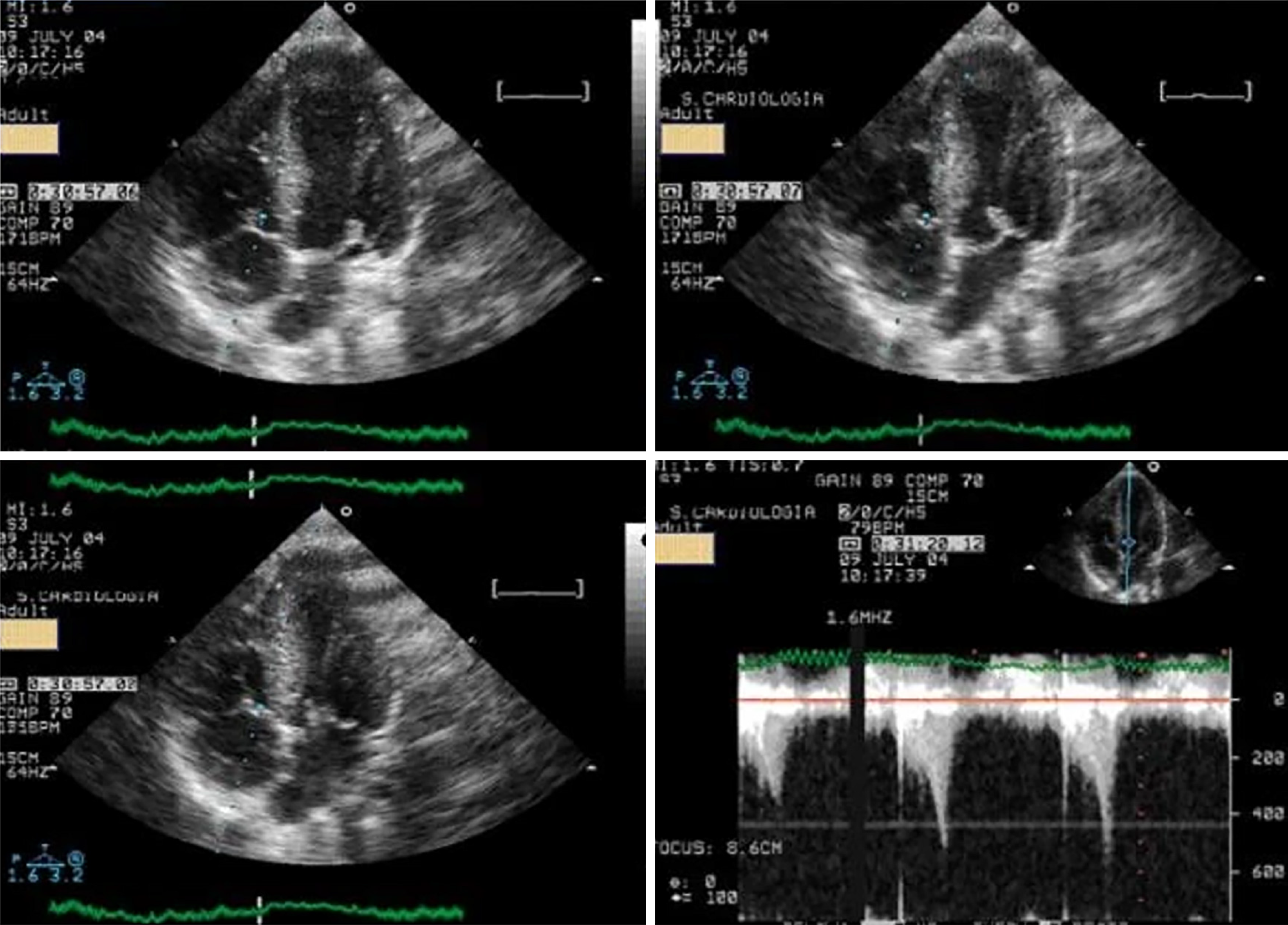
Figure 2 Systolic anterior movement of the mitral valve and significant intraventricular gradient detected at peak exercise.
Citation: Cotrim C, João I, Fazendas P, Almeida AR, Lopes L, Stuart B, Cruz I, Caldeira D, Loureiro MJ, Morgado G, Pereira H. Clinical applications of exercise stress echocardiography in the treadmill with upright evaluation during and after exercise. Cardiovasc Ultrasound 2013; 11: 26 [PMID: 23875614 DOI: 10.1186/1476-7120-11-26] Copyright © The Author (s) 2013. Published by BMC part of Springer Nature.
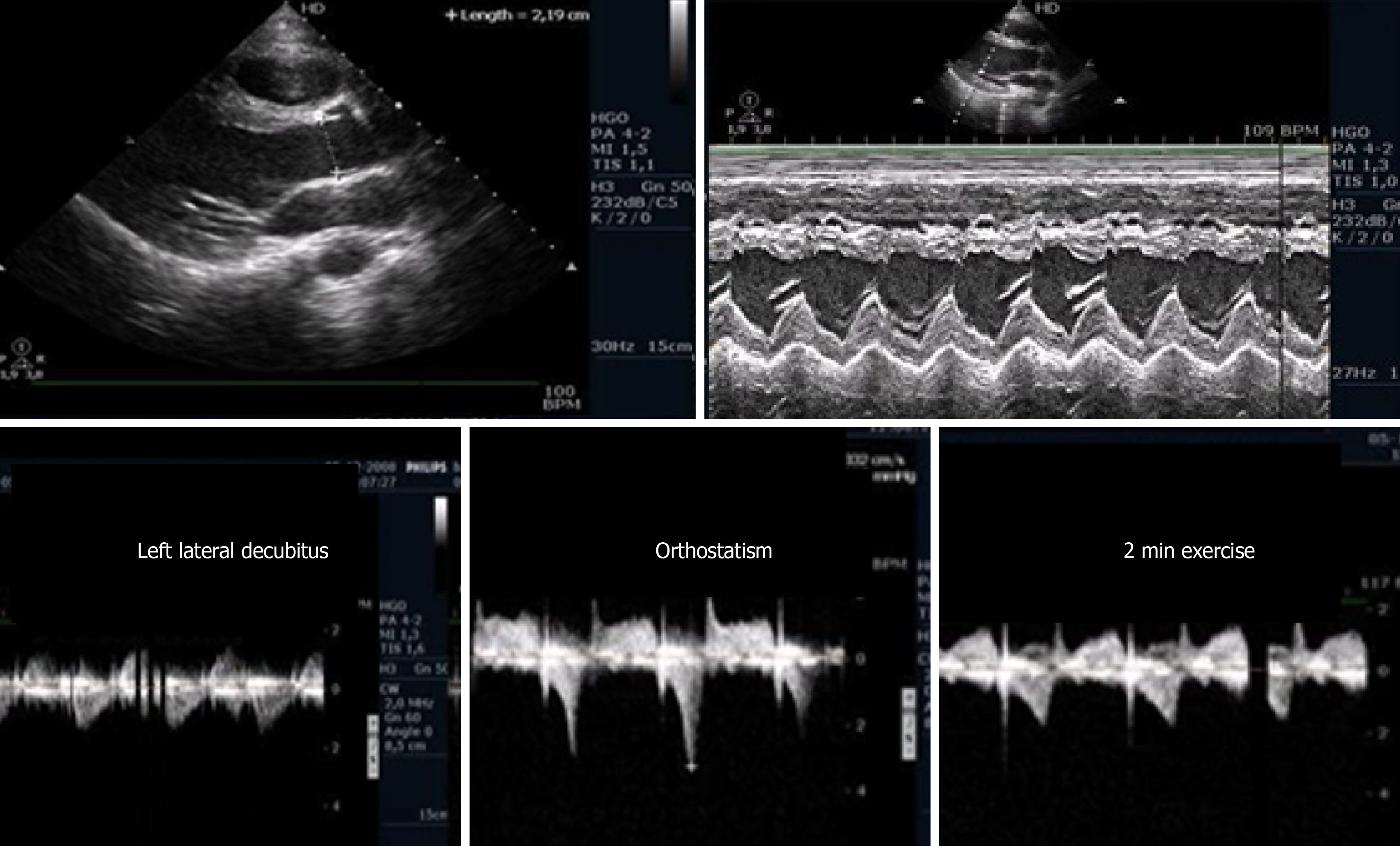
Figure 3 Intraventricular gradient present in the orthostatic position before exercise in an athlete, decreased during the initial phase of exercise testing.
Citation: Cotrim C, João I, Fazendas P, Almeida AR, Lopes L, Stuart B, Cruz I, Caldeira D, Loureiro MJ, Morgado G, Pereira H. Clinical applications of exercise stress echocardiography in the treadmill with upright evaluation during and after exercise. Cardiovasc Ultrasound 2013; 11: 26. [PMID: 23875614 DOI: 10.1186/1476-7120-11-26] Copyright © The Author (s) 2013. Published by BMC part of Springer Nature.
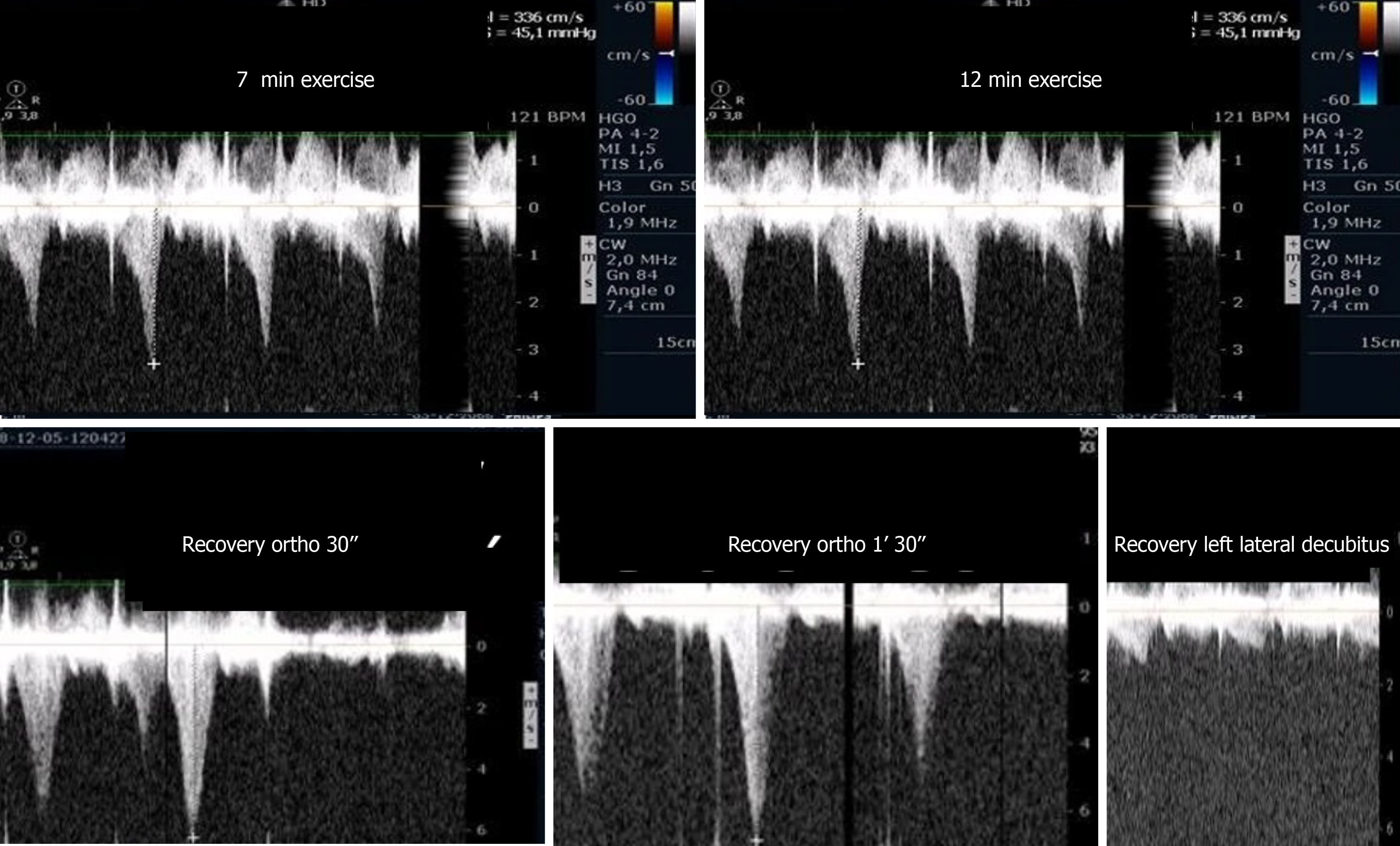
Figure 4 Intraventricular gradient increased during the last portion of the exercise test and after exercise in the orthostatic position.
Obstruction suddenly disappeared after placing the athlete in decubitus. Citation: Cotrim C, João I, Fazendas P, Almeida AR, Lopes L, Stuart B, Cruz I, Caldeira D, Loureiro MJ, Morgado G, Pereira H. Clinical applications of exercise stress echocardiography in the treadmill with upright evaluation during and after exercise. Cardiovasc Ultrasound 2013; 11: 26. [PMID: 23875614 DOI: 10.1186/1476-7120-11-26] Copyright © The Author (s) 2013. Published by BMC part of Springer Nature.
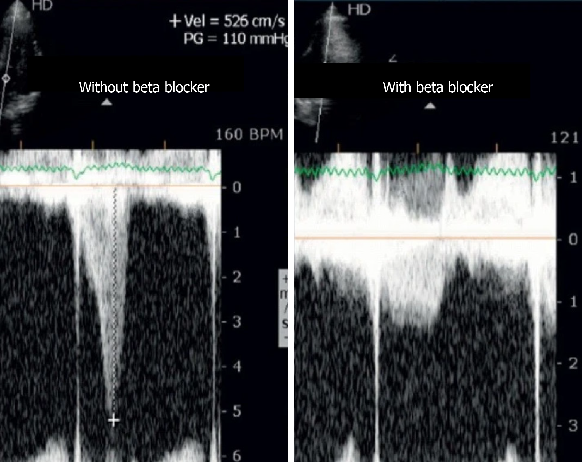
Figure 5 Intraventricular gradient in an athlete assessed before and on beta-blocker therapy.
Citation: Cotrim C, João I, Fazendas P, Almeida AR, Lopes L, Stuart B, Cruz I, Caldeira D, Loureiro MJ, Morgado G, Pereira H. Clinical applications of exercise stress echocardiography in the treadmill with upright evaluation during and after exercise. Cardiovasc Ultrasound 2013; 11: 26. [PMID: 23875614 DOI: 10.1186/1476-7120-11-26] Copyright © The Author (s) 2013. Published by BMC part of Springer Nature.
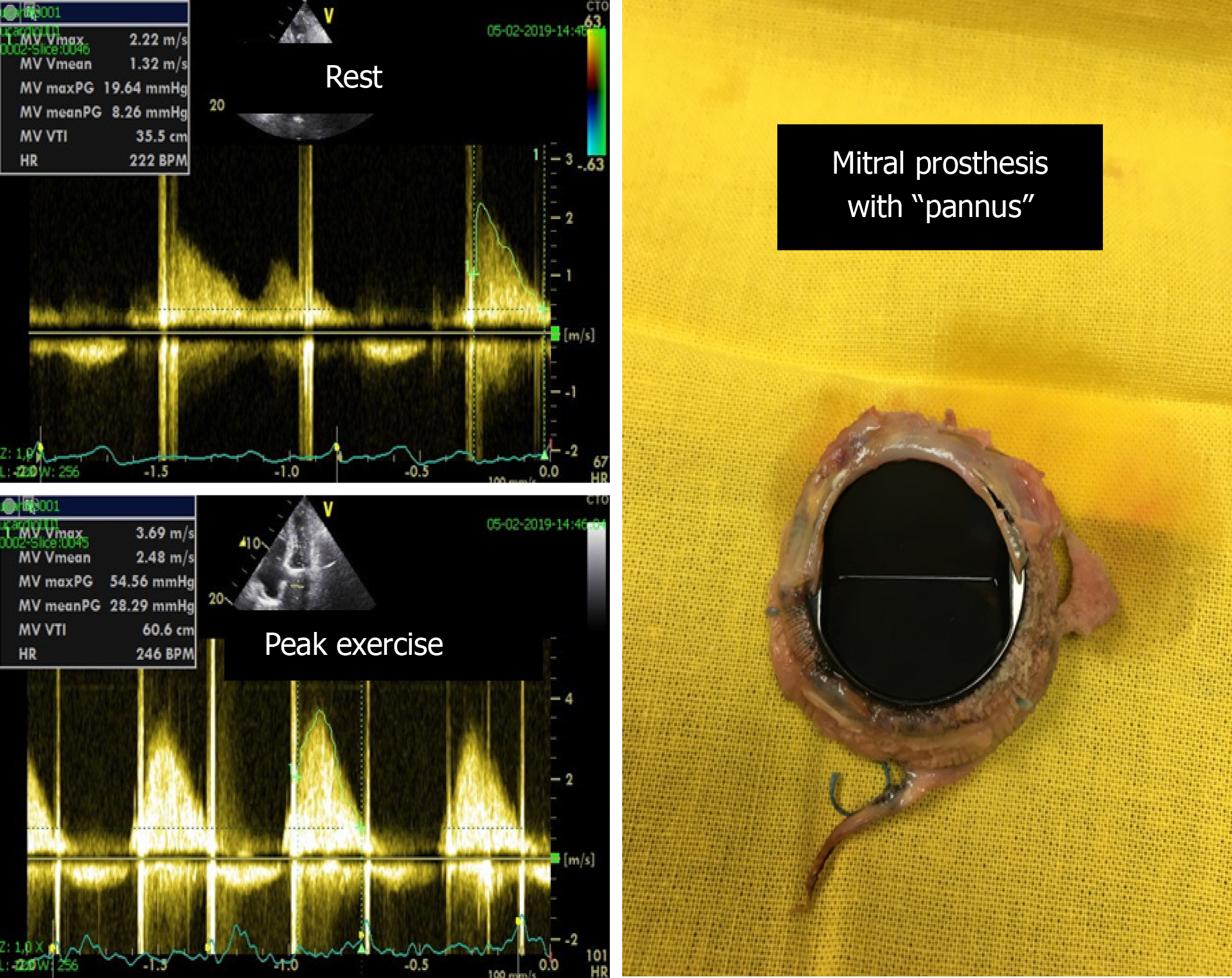
Figure 6 Significant increase in the mean gradient (from 8 mmHg to 28 mmHg) and appearance of severe symptoms with exercise in one patient with a mechanical mitral prosthesis with “pannus”.
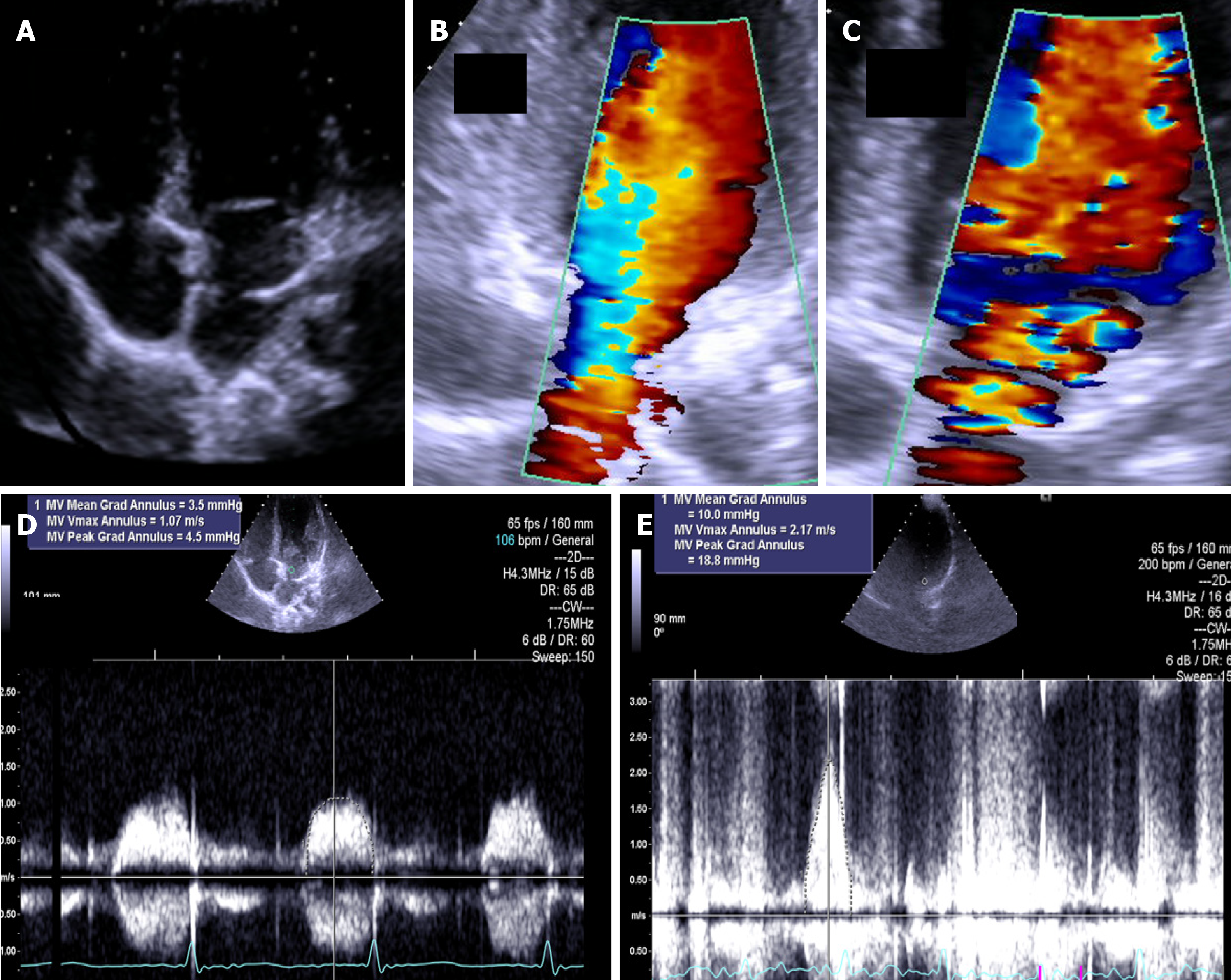
Figure 7 The exercise Doppler data in conjunction with the exercise data and clinical data led us to keep the patient in close clinical follow-up.
A: Intraauricular septum in “cor triatriatrium”; B: Color flow before exercise; C: Color flow at peak exercise; D: CW flow before exercise; E: CW flow at peak exercise.
Figure 8 Aortic gradient evaluated in a patient previously treated with a stent.
Based on the exercise stress echocardiography results, the patient was treated again.
- Citation: Cotrim CA, Café H, João I, Cotrim N, Guardado J, Cordeiro P, Cotrim H, Baquero L. Exercise stress echocardiography: Where are we now? World J Cardiol 2022; 14(2): 64-82
- URL: https://www.wjgnet.com/1949-8462/full/v14/i2/64.htm
- DOI: https://dx.doi.org/10.4330/wjc.v14.i2.64


