Copyright
©2014 Baishideng Publishing Group Inc.
World J Gastrointest Surg. Aug 27, 2014; 6(8): 164-168
Published online Aug 27, 2014. doi: 10.4240/wjgs.v6.i8.164
Published online Aug 27, 2014. doi: 10.4240/wjgs.v6.i8.164
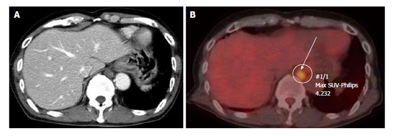
Figure 1 The tumor was 16 mm in size and located in the retrocrural space with suspicion of enlarged lymph nodes.
A: Computed tomography (CT) scan showing a 16-mm tumor in the retrocrural space of the lower mediastinum; B: Positron emission tomography-CT did not reveal any other abnormal accumulation. The mediastinal mass had 4.232 of the sum of the maximum standard uptake values. SUV: Standardized uptake value.
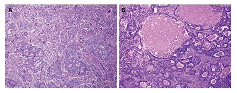
Figure 2 Histopathology revealed the widespread metastasis of moderately differentiated adenocarcinoma and necrosis.
Hematoxylin and eosin stain, A: × 10, B: × 50.
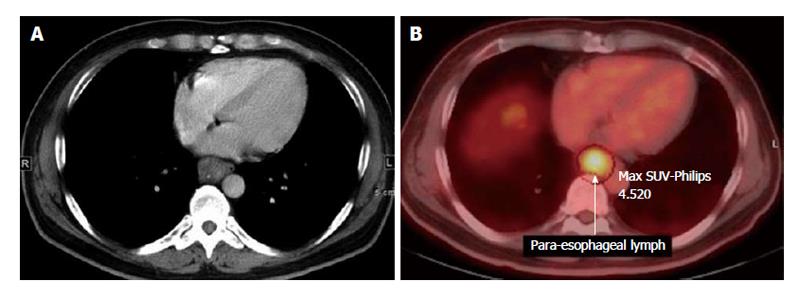
Figure 3 Computed tomography scan showing swelling of a para-esophageal lymph node (A), positron emission tomography-computed tomography did not reveal any other abnormal accumulation (B).
The mediastinal mass had 4.520 of the sum of the maximum standard uptake values. SUV: Standardized uptake value.
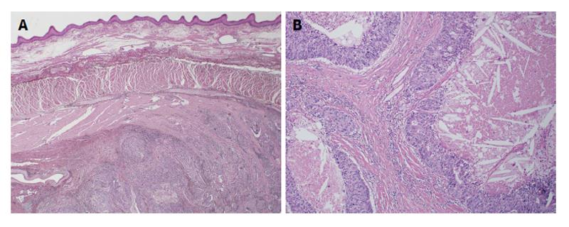
Figure 4 Histopathology showing no neoplastic change in the esophageal mucosal surface, compression from the esophageal adventitia, or infiltration of the muscularis propria.
Hematoxylin and eosin stain, A: × 10, B: × 50.
- Citation: Matsuda Y, Yano M, Miyoshi N, Noura S, Ohue M, Sugimura K, Motoori M, Kishi K, Fujiwara Y, Gotoh K, Marubashi S, Akita H, Takahashi H, Sakon M. Solitary mediastinal lymph node recurrence after curative resection of colon cancer. World J Gastrointest Surg 2014; 6(8): 164-168
- URL: https://www.wjgnet.com/1948-9366/full/v6/i8/164.htm
- DOI: https://dx.doi.org/10.4240/wjgs.v6.i8.164


