Copyright
©The Author(s) 2022.
World J Gastrointest Oncol. May 15, 2022; 14(5): 1057-1064
Published online May 15, 2022. doi: 10.4251/wjgo.v14.i5.1057
Published online May 15, 2022. doi: 10.4251/wjgo.v14.i5.1057
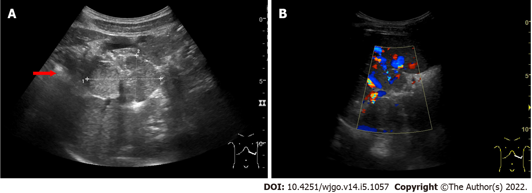
Figure 1 Ultrasound findings.
A: A well-defined, inhomogeneous echoic mass (red arrow) with a size of 5.9 cm × 3.8 cm was seen growing from the tail of the pancreas with both hyperechoic solid and hypoechoic cystic parts, dominated by a solid part; B: Color Doppler flow imaging showed dotted blood flow signals in the mass.
- Citation: Xu SY, Zhou B, Wei SM, Zhao YN, Yan S. Successful treatment of pancreatic accessory splenic hamartoma by laparoscopic spleen-preserving distal pancreatectomy: A case report. World J Gastrointest Oncol 2022; 14(5): 1057-1064
- URL: https://www.wjgnet.com/1948-5204/full/v14/i5/1057.htm
- DOI: https://dx.doi.org/10.4251/wjgo.v14.i5.1057


