Copyright
©The Author(s) 2015.
World J Gastrointest Oncol. Aug 15, 2015; 7(8): 95-101
Published online Aug 15, 2015. doi: 10.4251/wjgo.v7.i8.95
Published online Aug 15, 2015. doi: 10.4251/wjgo.v7.i8.95
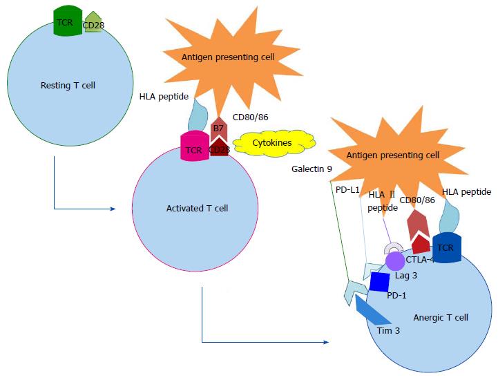
Figure 1 From a resting T cell to an activated or an anergic T cell.
To be activated a T cell lymphocyte needs recognition of an antigen coupled with major histocompatibility complex by its specific TCR, adequate cytokines and activation of co-stimulatory molecules such as CD28. An inhibitory signal can instead be transmitted by co-inhibitory molecules (PD-1, CTLA-4, Lag 3, Tim 3…) and lead to T cell anergy. TCR: T cell receptor; CD28: Cluster of differenciation 28; HLA: Human leucocyte antigen; CD80/86: Cluster of differenciation CD80/86; PD-1: Program death-1; PD-L1: Program death-ligand 1; CTLA-4: Cytotoxic T-lymphocyte-associated protein 4.
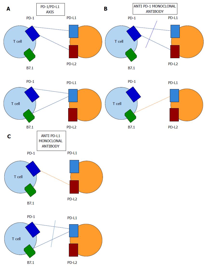
Figure 2 The program death-1 and program death-ligand 1 axis blockade.
A: The PD-1 and PD-L1 interactions: PD1 has two ligands called PD-L1 and PD-L2. PD-L1 can interact either with PD-1 or B7.1; B: Anti PD-1 monoclonal antibody blockade prevents PD-L1 and PD-L2 ligation to PD-1 but not the B7.1 and PD-L1 interaction; C: Anti PD-L1 monoclonal antibody blockade prevents PD-1 and B7.1 ligation to PD-L1 but not the PD-1 and PD-L2 interaction. PD-1: Program death-1; PD-L1: Program death-ligand 1; PD-L2: Program death-ligand 2.
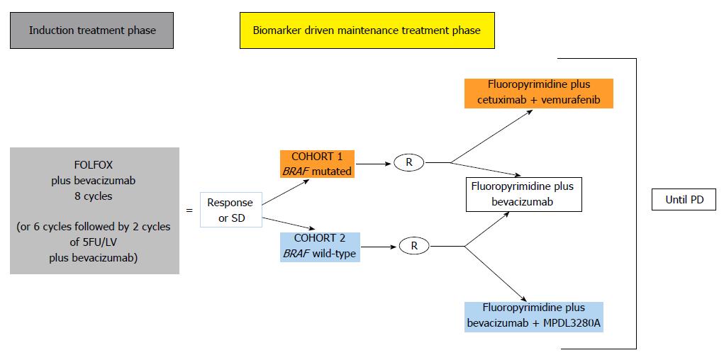
Figure 3 MODUL Phase III trial design.
5FU: 5-Fluoro-Uracil; LV: Leucovorin; SD: Stable disease; R: Randomization; PD: Progressive disease.
- Citation: Guillebon E, Roussille P, Frouin E, Tougeron D. Anti program death-1/anti program death-ligand 1 in digestive cancers. World J Gastrointest Oncol 2015; 7(8): 95-101
- URL: https://www.wjgnet.com/1948-5204/full/v7/i8/95.htm
- DOI: https://dx.doi.org/10.4251/wjgo.v7.i8.95


