Copyright
©2013 Baishideng Publishing Group Co.
World J Gastrointest Oncol. Jul 15, 2013; 5(7): 159-170
Published online Jul 15, 2013. doi: 10.4251/wjgo.v5.i7.159
Published online Jul 15, 2013. doi: 10.4251/wjgo.v5.i7.159
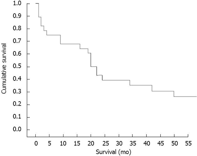
Figure 1 Survival curve of a series of 29 patients with resected hilar cholangiocarcinoma.
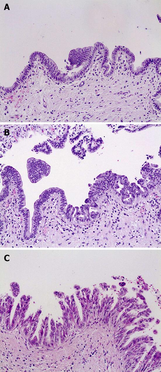
Figure 2 Biliary intraepithelial neoplasia.
A: Biliary intraepithelial neoplasia (BilIN) 1 (HE stain, ×200); B: BilIN 2 (HE stain, ×100); C: BilIN 3 (HE stain, ×100).
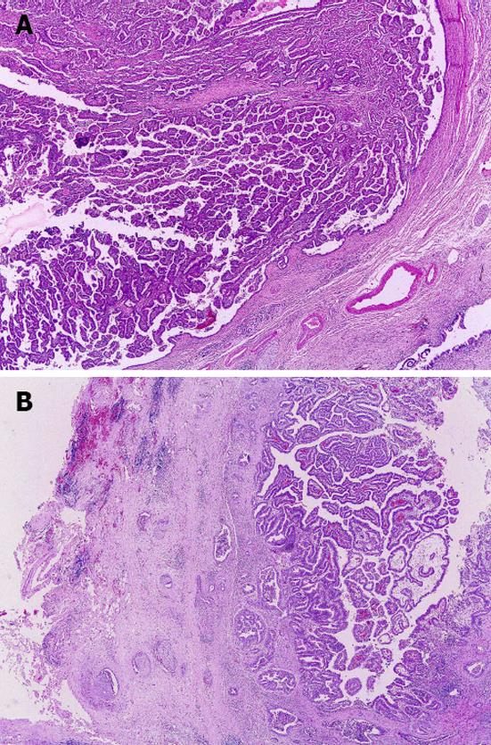
Figure 3 Intraductal papillary neoplasm of the biliary tract.
A: Intraductal papillary neoplasm of the biliary tract (IPNB) without invasion, classically named biliary papillomatosis (HE stain, ×20); B: IPNB with associated invasive carcinoma, previously named invasive papillary carcinoma (HE stain, × 20).
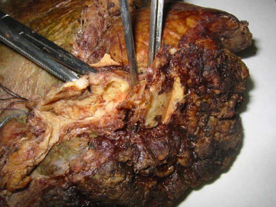
Figure 4 An example of a nodular-sclerosing cholangiocarcinoma in the common, right and left hepatic ducts.
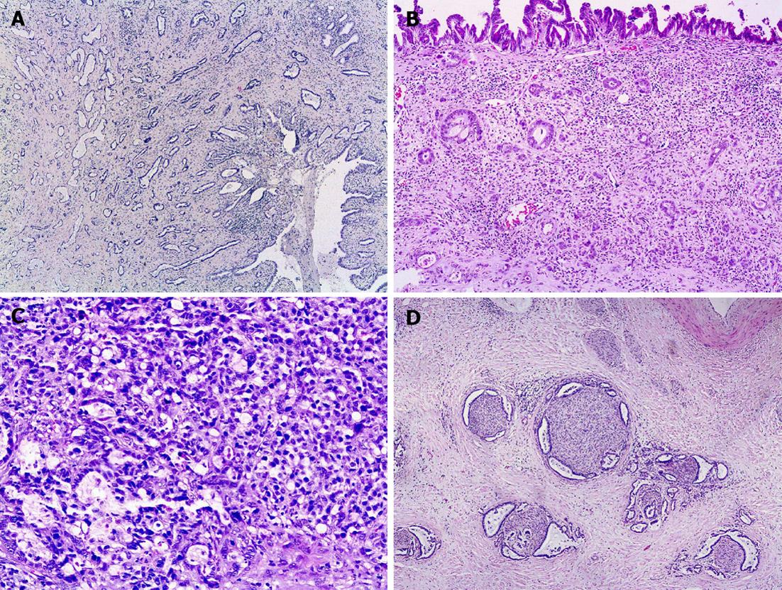
Figure 5 Examples of the broad morphological spectrum of hilar bile duct carcinoma.
A: Well differentiated biliary type adenocarcinoma (HE stain, ×40); B: A case with well defined tumor glands interspersed with poorly differentiated small tumor groups an single tumor cells (besides, biliary intraepithelial neoplasia 3 can be observed on the duct surface) (HE stain, ×100); C: A case with poorly differentiated cell component with resembling signet ring cells (HE stain, ×200); D: Perineural invasion by well differentiated glands (HE stain, ×40).
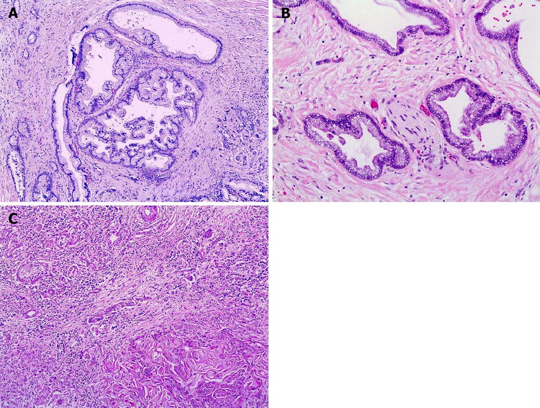
Figure 6 Histological variants.
A: Gastric foveolar type carcinoma (HE stain, ×100); B: Very well differentiated glands of a gastric foveolar type carcinoma near a nervous fascicle (HE stain, ×200); C: Adenosquamous carcinoma showing the glandular and squamous component (HE stain, ×100).
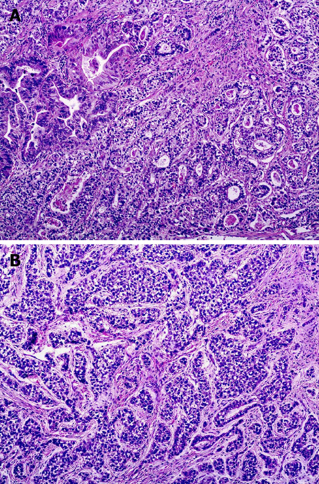
Figure 7 Histological variants: clear cell carcinoma (HE stain, ×100).
A: An area with glandular pattern; B: An area with trabecular pattern.
- Citation: Castellano-Megías VM, Ibarrola-de Andrés C, Colina-Ruizdelgado F. Pathological aspects of so called "hilar cholangiocarcinoma". World J Gastrointest Oncol 2013; 5(7): 159-170
- URL: https://www.wjgnet.com/1948-5204/full/v5/i7/159.htm
- DOI: https://dx.doi.org/10.4251/wjgo.v5.i7.159


