Copyright
©2011 Baishideng Publishing Group Co.
World J Gastrointest Oncol. Jul 15, 2011; 3(7): 116-118
Published online Jul 15, 2011. doi: 10.4251/wjgo.v3.i7.116
Published online Jul 15, 2011. doi: 10.4251/wjgo.v3.i7.116
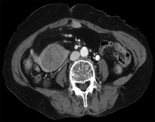
Figure 1 A computed tomography scan demonstrating a fatty mass with a solid portion.
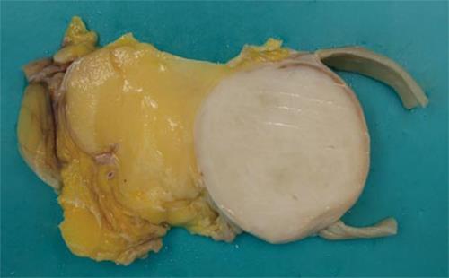
Figure 2 Gross picture showing one portion of a whitish, elastic-hard nodule located in the small bowel wall and the other portion of yellow, soft nodule located in the small bowel mesentery.
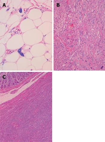
Figure 3 Histological examinations of different areas in the mass.
A: Well-differentiated component showing atypical cells with nucleomegaly and hyperchromasia scattered within mature adipocytes (HE stain, × 400); B: The dedifferentiated component showing pleomorphic spindle cells arranged in a fascicular pattern (HE stain, × 200); C: The dedifferentiated component invading into the bowel wall (HE stain, × 40).
- Citation: Cha EJ. Dedifferentiated liposarcoma of the small bowel mesentery presenting as a submucosal mass. World J Gastrointest Oncol 2011; 3(7): 116-118
- URL: https://www.wjgnet.com/1948-5204/full/v3/i7/116.htm
- DOI: https://dx.doi.org/10.4251/wjgo.v3.i7.116


