Copyright
©2010 Baishideng.
World J Gastrointest Oncol. Feb 15, 2010; 2(2): 121-124
Published online Feb 15, 2010. doi: 10.4251/wjgo.v2.i2.121
Published online Feb 15, 2010. doi: 10.4251/wjgo.v2.i2.121
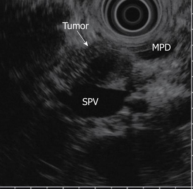
Figure 1 EUS image of pancreatic GCT.
The tumor showed a homogeneous pattern and regular borders (arrow). EUS: Endoscopic ultrasonography; GCT: Granular cell tumor; MPD: main pancreatic duct; PV: portal vein.
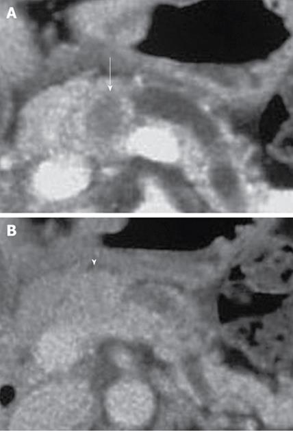
Figure 2 CT image of pancreatic GCT.
A: CT showing poor enhancement of the tumor compared with that of the surrounding pancreatic parenchyma at early phase o dynamic CT (arrow); B: CT showing gradual enhancement of the tumor at delayed phase (arrowhead). CT: Computed tomography.
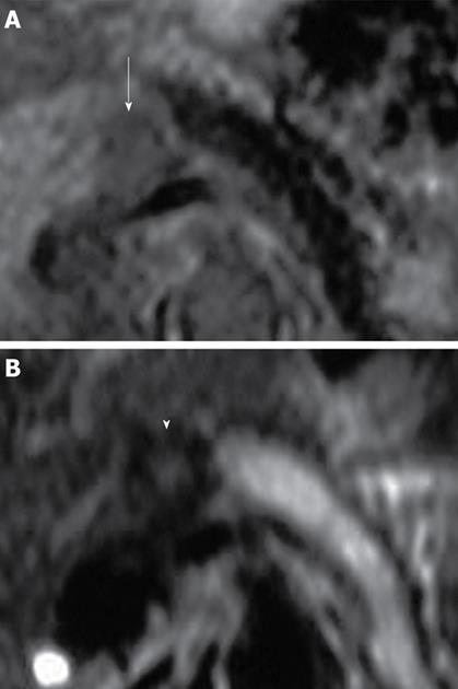
Figure 3 MRI findings of pancreatic GCT.
A: The tumor showed a hypointense mass in the T1-weighted image (arrow); B: The surrounding and center of the tumor were hypointense and hyperintense on T2-weighted image, respectively (arrowhead). MRI: magnetic resonance imaging.
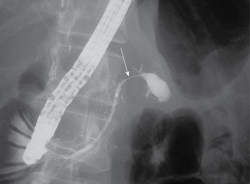
Figure 4 ERCP showing the stricture and the dilatation in the distal pancreatic duct (arrow).
ERCP: Endoscopic retrograde cholangiopancreatography.
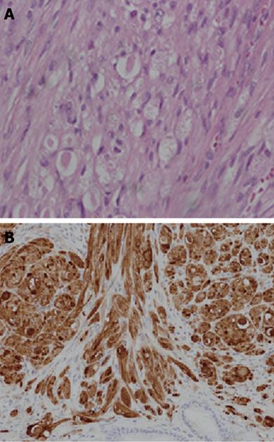
Figure 5 Histological findings.
A: Microscopic study showed a well-limited nodule made of large clusters of benign cells with small nuclei and abundant granular cytoplasm; B: S-100 protein staining was positive in the cell cytoplasm.
- Citation: Kanno A, Satoh K, Hirota M, Hamada S, Umino J, Itoh H, Masamune A, Egawa S, Motoi F, Unno M, Ishida K, Shimosegawa T. Granular cell tumor of the pancreas: A case report and review of literature. World J Gastrointest Oncol 2010; 2(2): 121-124
- URL: https://www.wjgnet.com/1948-5204/full/v2/i2/121.htm
- DOI: https://dx.doi.org/10.4251/wjgo.v2.i2.121


