Copyright
©The Author(s) 2025.
World J Gastrointest Oncol. Mar 15, 2025; 17(3): 99304
Published online Mar 15, 2025. doi: 10.4251/wjgo.v17.i3.99304
Published online Mar 15, 2025. doi: 10.4251/wjgo.v17.i3.99304
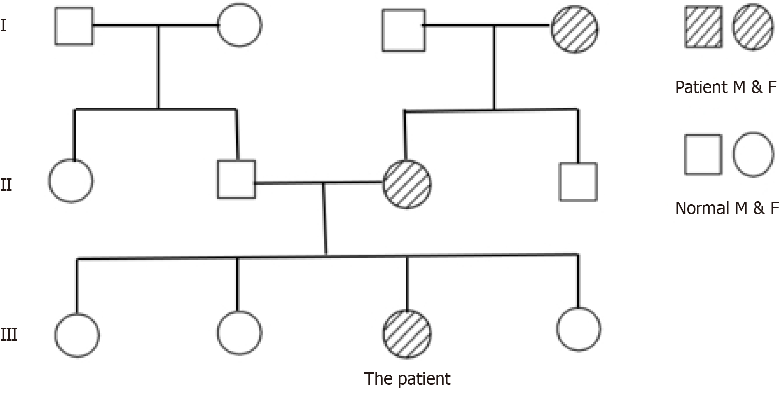
Figure 1
Family history of the patient.
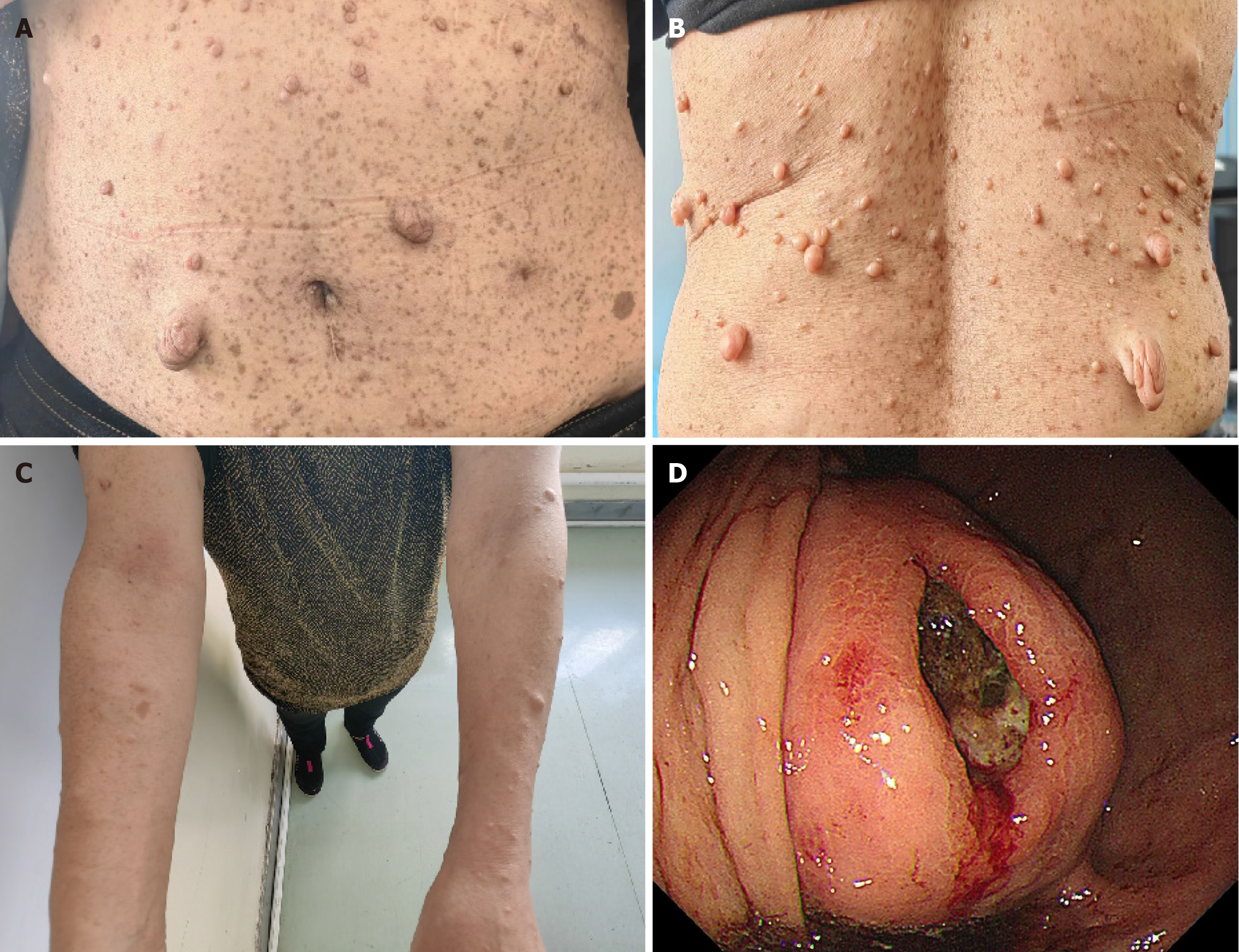
Figure 2 Clinical presentation of the patient.
A: Multiple café-au-lait patches and cutaneous nodular masses on the abdomen; B: Multiple café-au-lait patches and cutaneous nodular masses on the back; C: Multiple café-au-lait patches and cutaneous nodular masses on the arm; D: Gastric stromal tumor with bleeding ulcer in gastroscopy.
Figure 3 Imaging manifestations of the patient.
A: Huge stromal tumor in the abdominal cavity, squeezing abdominal organs; B: Cutaneous nodular masse on the abdomen; C: Subcutaneous nodule masse on the back; D: 12 months after surgery, no relapse in the tangential line and abdominal cavity.
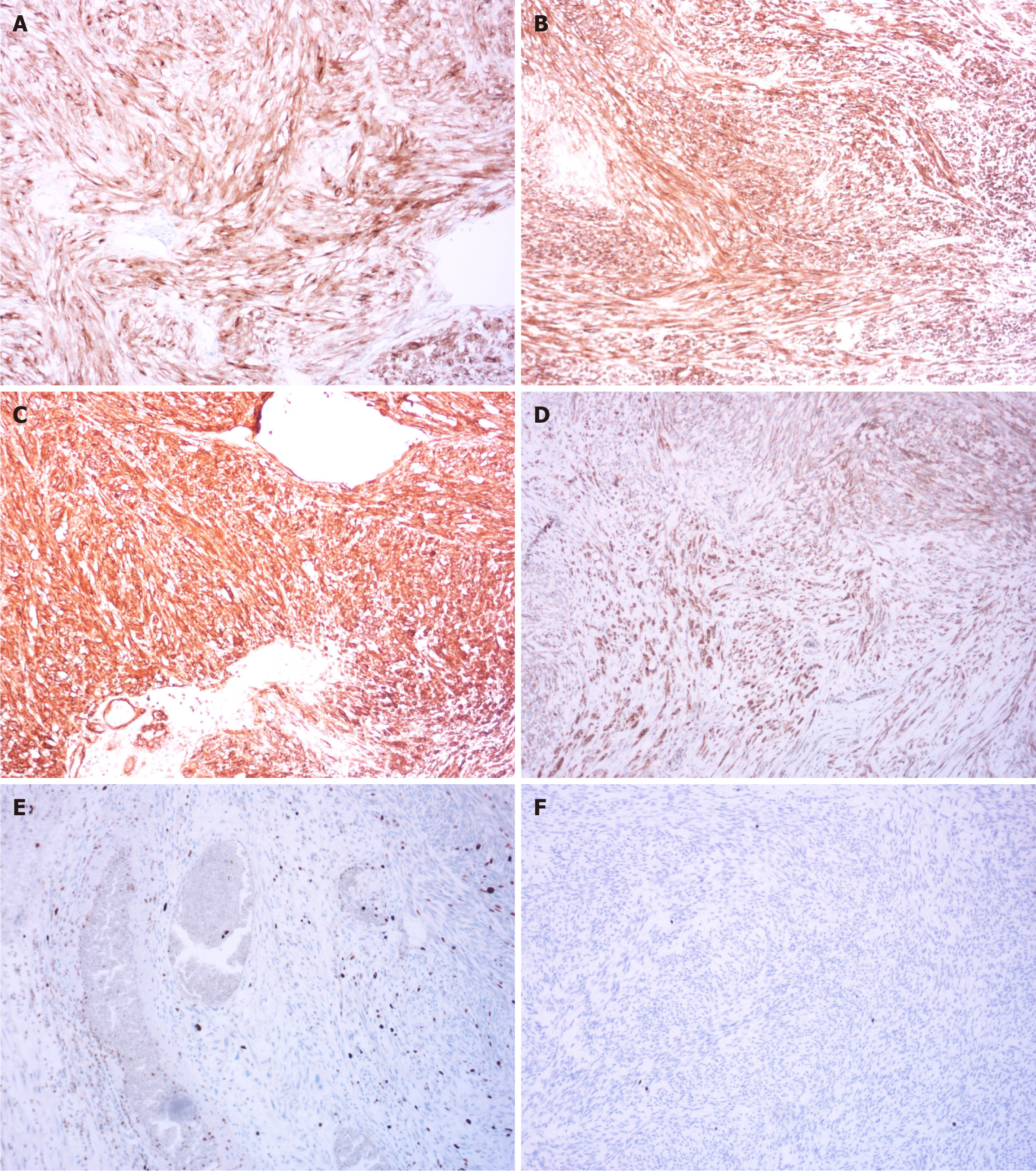
Figure 4 Postoperative immunohistochemical results.
A: Diffusely strong expression of CD117 (× 100); B: Diffusely strong expression of delay of germination 1 (× 100); C: Diffusely strong expression of CD34 (× 100); D: Diffusely strong expression of succinate dehydrogenase complex iron sulfur subunit B (× 100); E: Diffusely strong expression of Ki67 (× 100); F: Diffusely strong expression of phosphorylated histone H3 (× 100).
Figure 5
Mutation site on chromosome.
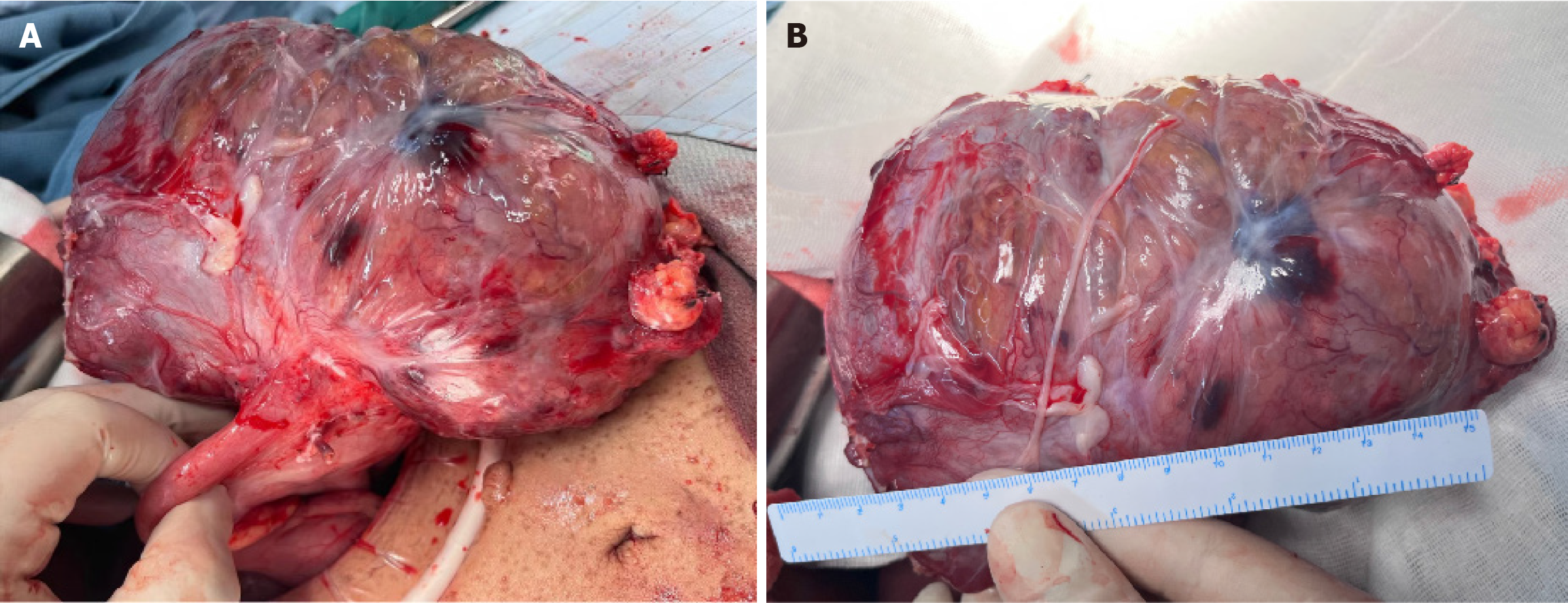
Figure 6 Intraoperative view.
A: The stromal tumor is from the stomach; B: Size of stromal tumor.
- Citation: Bai GY, Shan KS, Li CS, Wang XH, Feng MY, Gao Y. Gastric gastrointestinal stromal tumor in a patient with neurofibromatosis type I presenting with anemia: A case report. World J Gastrointest Oncol 2025; 17(3): 99304
- URL: https://www.wjgnet.com/1948-5204/full/v17/i3/99304.htm
- DOI: https://dx.doi.org/10.4251/wjgo.v17.i3.99304


