Copyright
©The Author(s) 2025.
World J Gastrointest Oncol. Mar 15, 2025; 17(3): 101991
Published online Mar 15, 2025. doi: 10.4251/wjgo.v17.i3.101991
Published online Mar 15, 2025. doi: 10.4251/wjgo.v17.i3.101991
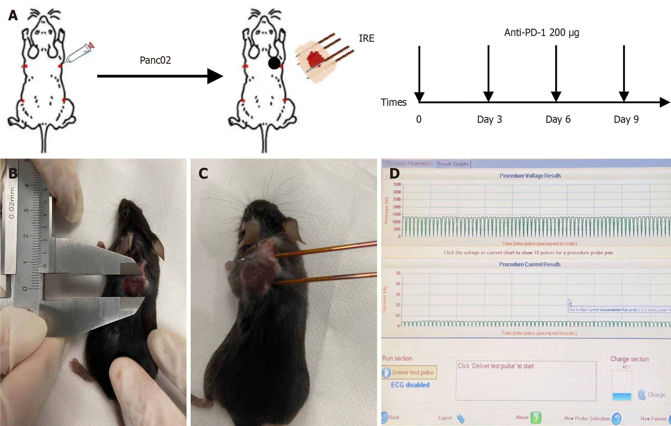
Figure 1 Establishment of pancreatic cancer model and ablation parameters.
A: Schematic drawing of the study design. C57BL/6 mice were enrolled for treatment once the tumor size reached about 10 mm; B: Tumor growth on day 20; C: Irreversible electroporation procedure was performed with two electrodes; D: Ablation parameters (1200 kV/cm, 70 pulses, 70 μs). IRE: Irreversible electroporation; PD-1: Programmed cell death protein 1.
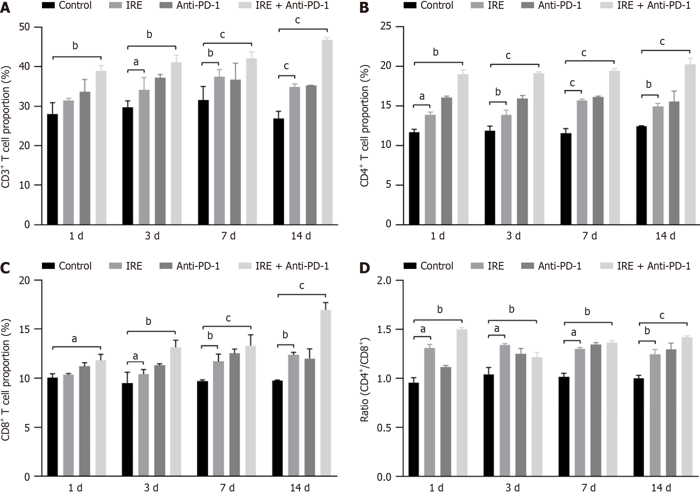
Figure 2 Irreversible electroporation combined with anti-programmed cell death protein 1 treatment increased T lymphocyte infiltration in peripheral blood cells.
A: Percentages of CD3+ T cells within a single-cell suspension of tumor tissues; B: Percentages of CD4+ T cells; C: Percentages of CD8+ T cells; D: CD4+: CD8+ ratio (values represent mean ± standard deviation, aP < 0.05, bP < 0.01, cP < 0.001). IRE: Irreversible electroporation; PD-1: Programmed cell death protein 1.
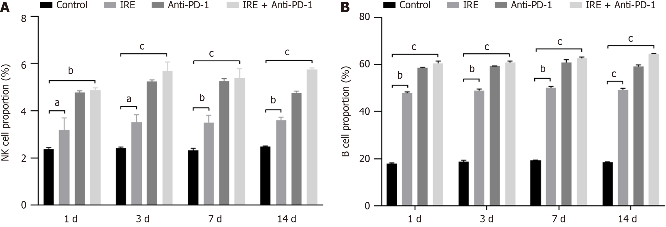
Figure 3 Irreversible electroporation combined with anti-programmed cell death protein 1 treatment increased peripheral blood cell infiltration of natural killer and B cells.
A: Percentages of natural killer cells; B: Percentages of B cells (values represent mean ± standard deviation, aP < 0.05, bP < 0.01, cP < 0.001). IRE: Irreversible electroporation; NK: Natural killer; PD-1: Programmed cell death protein 1.
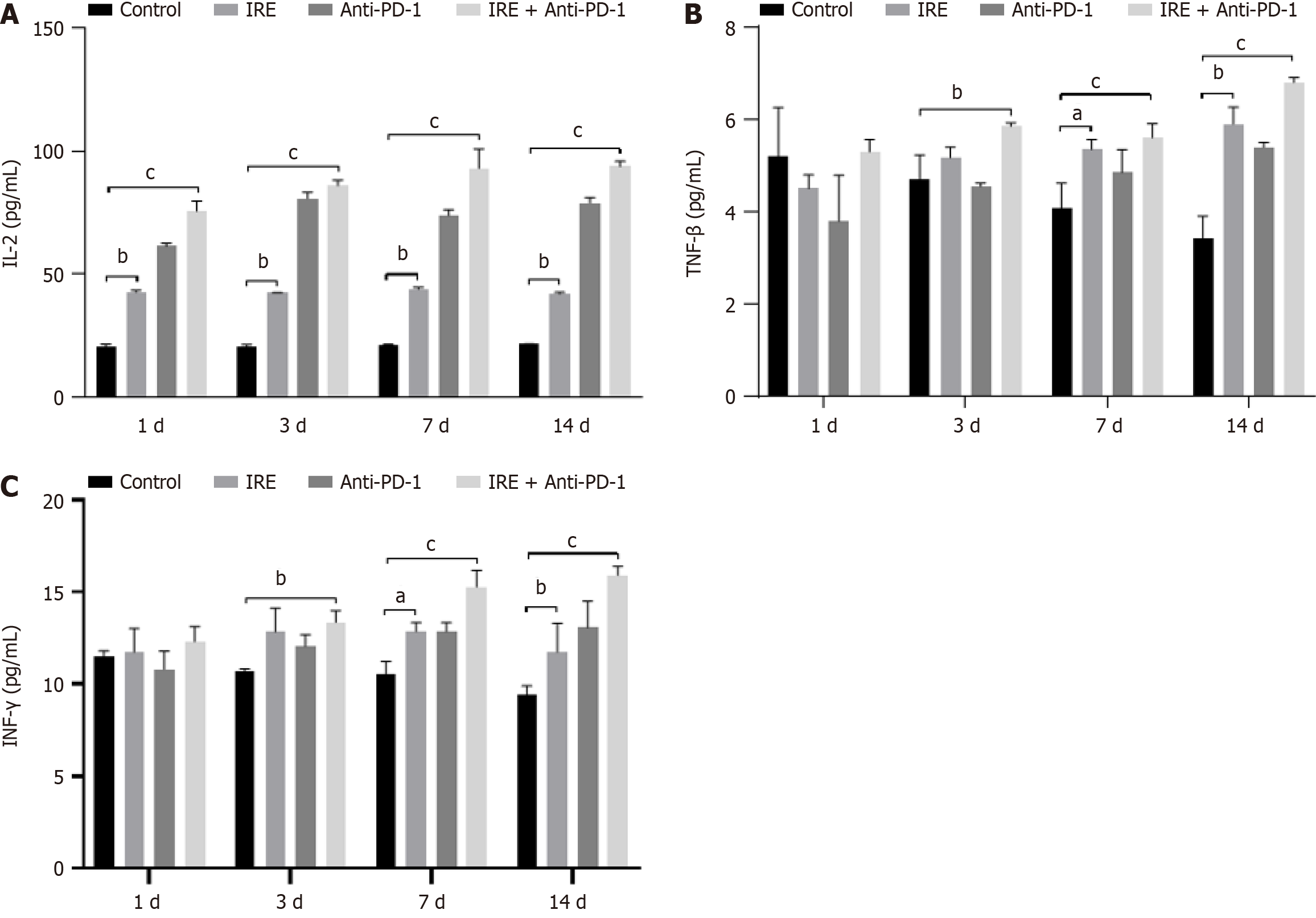
Figure 4 Irreversible electroporation combined programmed cell death protein 1 treatment modulated the concentrations of T helper type 1 cytokines in the peripheral blood.
A: Plasma concentration of interleukin-2; B: Plasma concentration of tumor necrosis factor-β; C: Plasma concentration of interferon-γ (values represent mean ± standard deviation, aP < 0.05, bP < 0.01, cP < 0.001). IL-2: Interleukin-2; INF-γ: Interferon-γ; IRE: Irreversible electroporation; PD-1: Programmed cell death protein 1; TNF-β: Tumor necrosis factor-β.
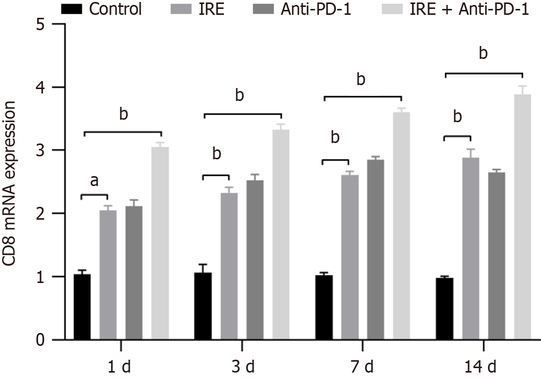
Figure 5 Irreversible electroporation combined programmed cell death protein 1 treatment increased the mRNA expression of CD8 in the tumor tissue.
Values represent mean ± standard deviation. aP < 0.01, bP < 0.001. IRE: Irreversible electroporation; PD-1: Programmed cell death protein 1. IRE: Irreversible electroporation; PD-1: Programmed cell death protein 1.
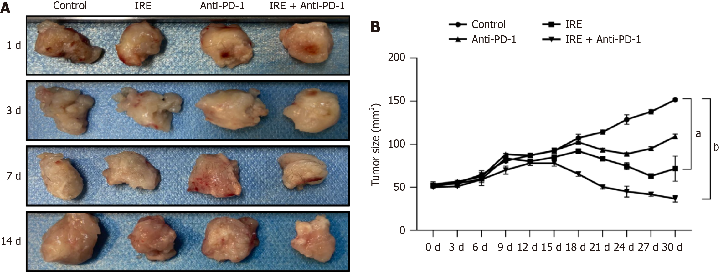
Figure 6 Irreversible electroporation combined programmed cell death protein 1 treatment increased the tumor regression.
A: Tumor morphologic changes; B: Tumor size changes (values represent mean ± standard deviation, aP < 0.01, bP < 0.001). IRE: Irreversible electroporation; PD-1: Programmed cell death protein 1.
- Citation: Ma YY, Wang XH, Zeng JY, Chen JB, Niu LZ. Irreversible electroporation combined with anti-programmed cell death protein 1 therapy promotes tumor antigen-specific CD8+ T cell response. World J Gastrointest Oncol 2025; 17(3): 101991
- URL: https://www.wjgnet.com/1948-5204/full/v17/i3/101991.htm
- DOI: https://dx.doi.org/10.4251/wjgo.v17.i3.101991


