Copyright
©The Author(s) 2024.
World J Gastrointest Oncol. Aug 15, 2024; 16(8): 3529-3538
Published online Aug 15, 2024. doi: 10.4251/wjgo.v16.i8.3529
Published online Aug 15, 2024. doi: 10.4251/wjgo.v16.i8.3529
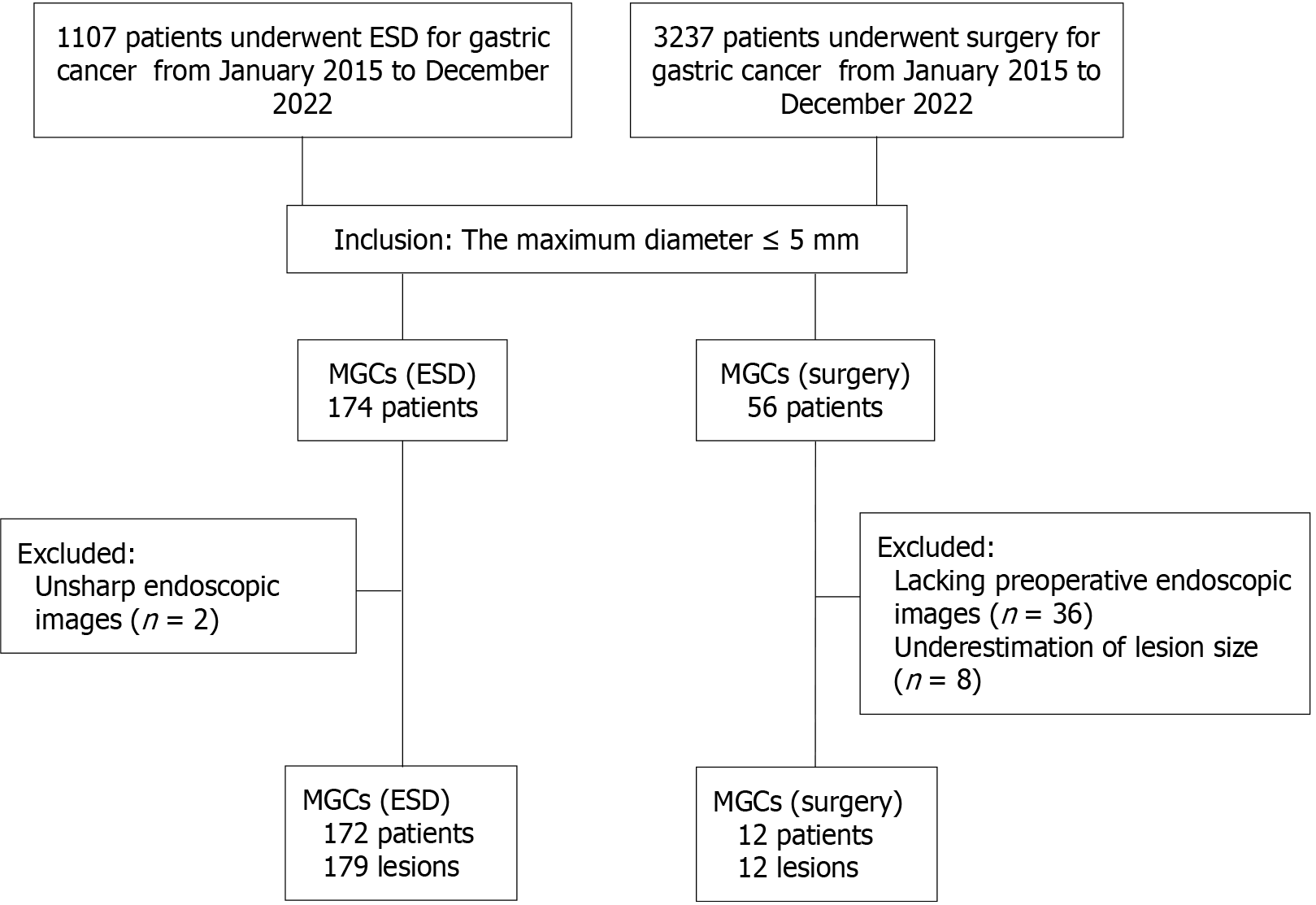
Figure 1 Flowchart of patient enrollment.
MGCs: Minute gastric cancers; ESD: Endoscopic submucosal dissection.
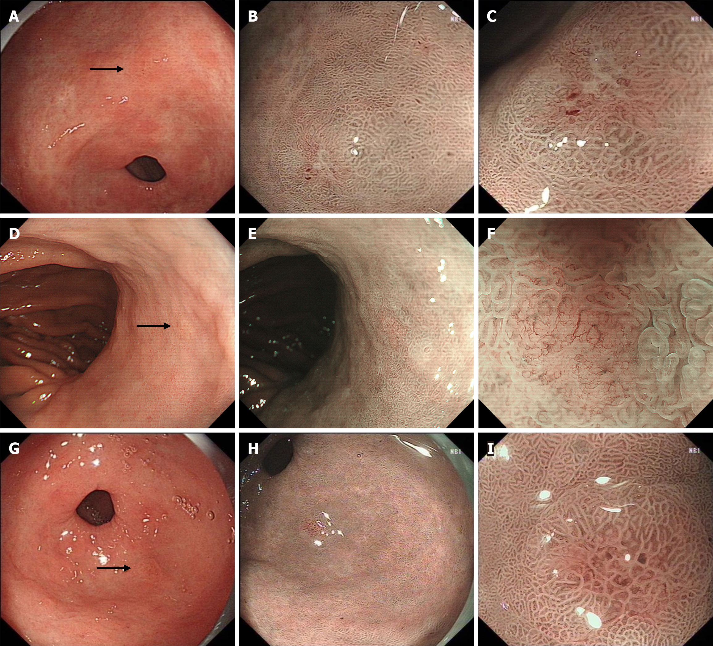
Figure 2 Minute gastric cancers found by narrow-band imaging.
A–C: 0-IIc lesion in the posterior wall of gastric antrum; D–F: 0-IIb lesion in lesser curvature of gastric body; G–I: 0-IIa lesion in the greater curvature of gastric antrum; A, D and G: Lesions were difficult to be detected under white light endoscopy (arrow); B, E and H: After switching to narrow-band imaging (NBI) endoscopy, the lesions were brownish under NBI; C, F and I: Irregular microstructures and/or microvessels were seen under magnifying endoscopy with NBI.
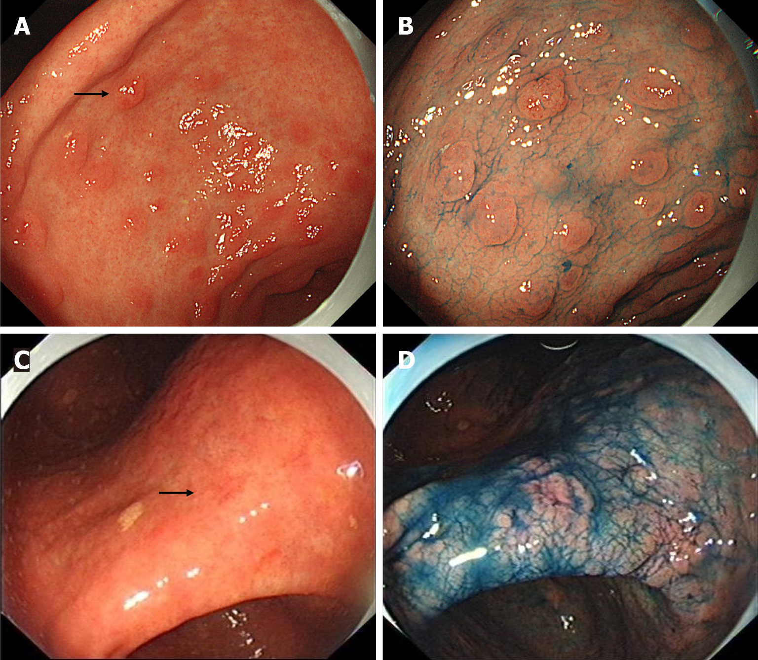
Figure 3 Minute gastric cancers found by indigo carmine.
A and B: 0-IIa lesion near the gastric angle; C and D: 0-IIc lesion in the gastric angle. A and C: Lesions were difficult to be detected under white light endoscopy (arrow); B and D: After spraying indigo carmine, the lesions were more obvious.
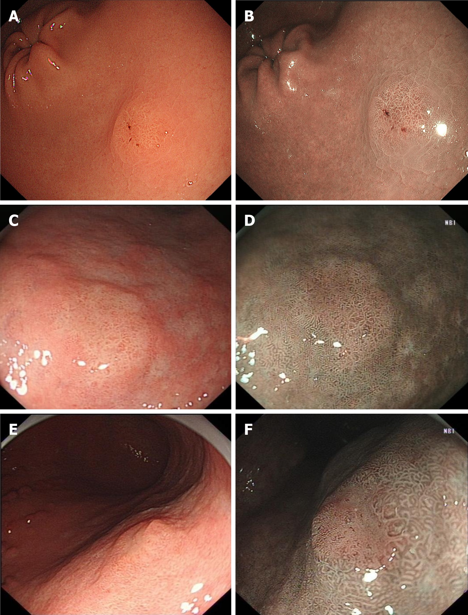
Figure 4 Minute gastric cancers diagnosed only using white light endoscopy combined with narrow-band imaging.
A and B: 0-IIa lesion in the posterior wall of the gastric antrum; C and D: 0-IIa lesion in lesser curvature of the gastric body; E and F: 0-IIa lesion in the posterior wall of the gastric body. A, C and E: Lesions were yellowish-red or whitish under white light endoscopy; B, D and F: Lesions were brownish under narrow-band imaging with a clear demarcation line.
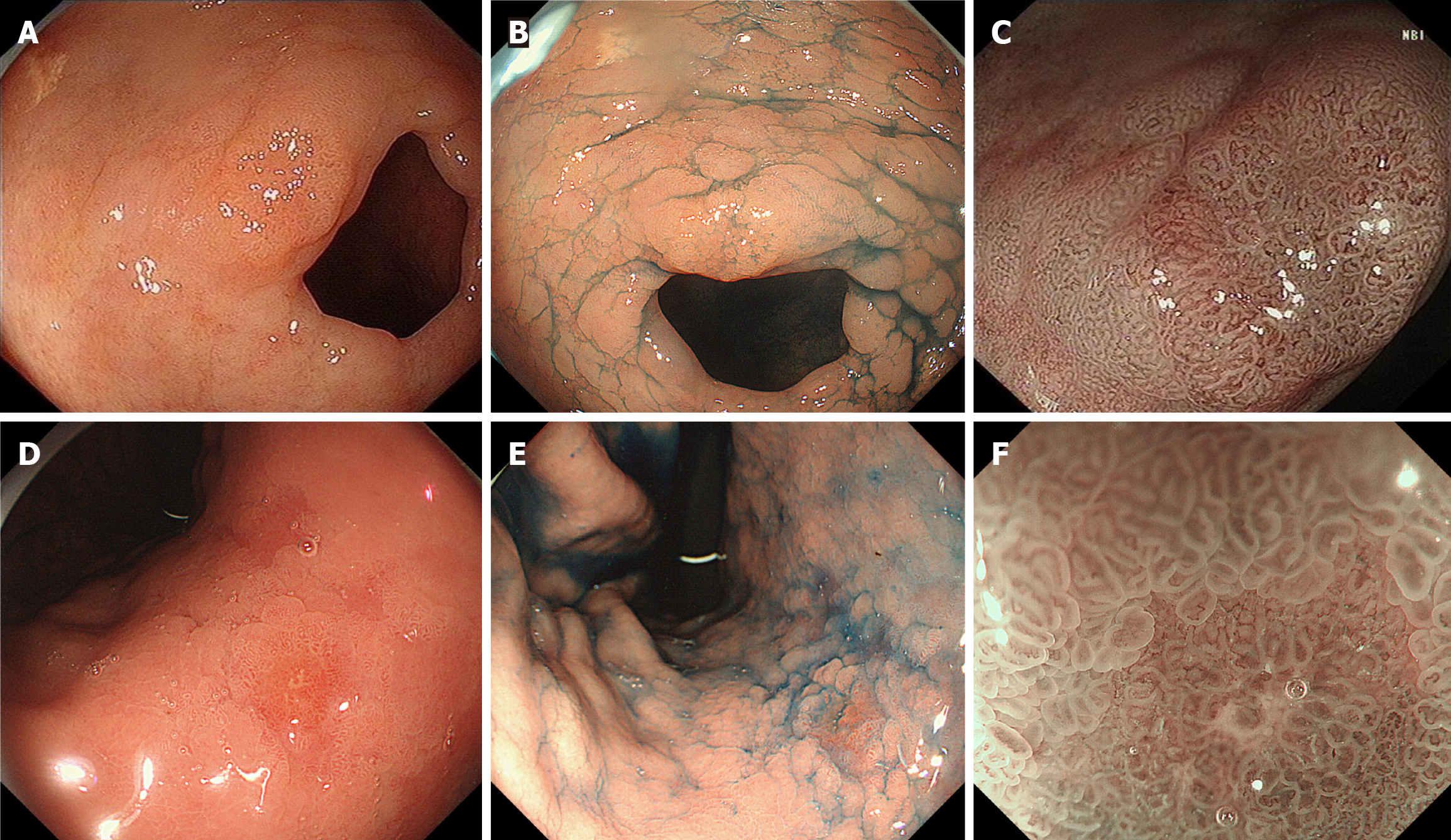
Figure 5 Diagnostic differences between high-confidence and low-confidence diagnosis groups using magnifying endoscopy with narrow-band imaging.
A-C: 0-IIa lesion in the lesser curvature of the pyloric duct. Magnifying endoscopy with narrow-band imaging (ME-NBI) showed irregular microstructure and microvessels with a clear demarcation line, classified as high confidence in endoscopic diagnosis; D-F: 0-IIc type lesion in the lesser curvature of the gastric body. ME-NBI showed regular microstructure and slightly thickened microvessels, classified as low confidence in endoscopic diagnosis.
- Citation: Ji XW, Lin J, Wang YT, Ruan JJ, Xu JH, Song K, Mao JS. Endoscopic detection and diagnostic strategies for minute gastric cancer: A real-world observational study. World J Gastrointest Oncol 2024; 16(8): 3529-3538
- URL: https://www.wjgnet.com/1948-5204/full/v16/i8/3529.htm
- DOI: https://dx.doi.org/10.4251/wjgo.v16.i8.3529


