Copyright
©The Author(s) 2024.
World J Gastrointest Oncol. Jul 15, 2024; 16(7): 3230-3240
Published online Jul 15, 2024. doi: 10.4251/wjgo.v16.i7.3230
Published online Jul 15, 2024. doi: 10.4251/wjgo.v16.i7.3230
Figure 1 Identifying high expression of aldehyde dehydrogenase 2 family member in colorectal cancer cells.
A: Western blot analysis of aldehyde dehydrogenase 2 family member (ALDH2) protein expression in colorectal cancer (CRC) cell lines (CL-40, SK-CO-1, SW-403, HT-29, COLO-678, and SW480) and human normal colon epithelial cell line (NCM460); B: Quantitative reverse transcriptase PCR analysis of ALDH2 expression in human CRC cell lines as indicated. Data are displayed as the mean ± SD. aP < 0.05, bP < 0.01, cP < 0.001. ALDH2: Aldehyde dehydrogenase 2 family member.
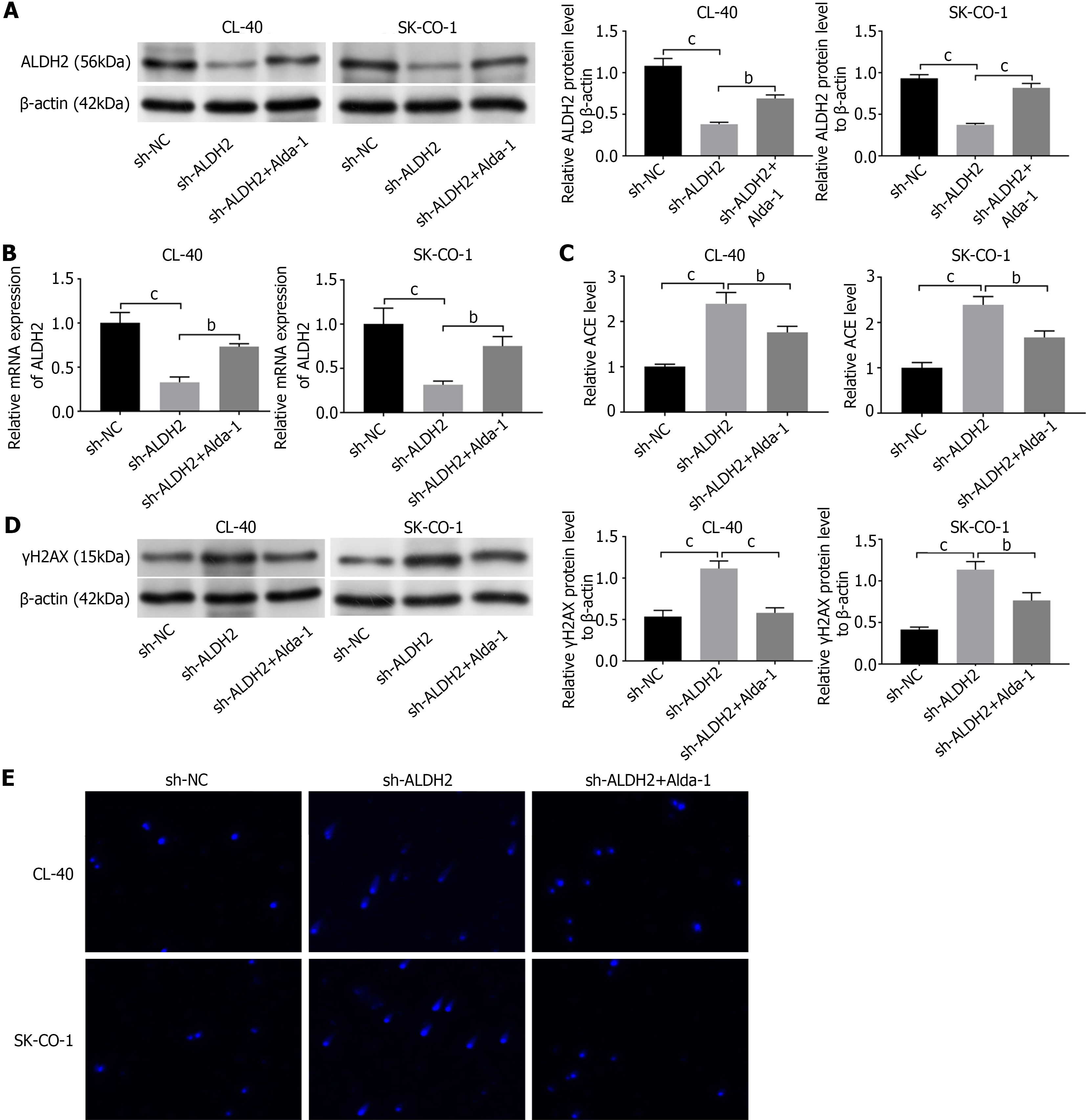
Figure 2 Aldehyde dehydrogenase 2 family member promotes the accumulated acetaldehyde and DNA damage of colorectal cancer cells.
A: Western blot measured the transfection efficiency of shRNA aldehyde dehydrogenase 2 family member (sh-ALDH2) and treated with Alda-1 (1 μM) in CL-40 and SK-CO-1 cells; B: Quantitative reverse transcriptase PCR measured the transfection efficiency of sh-ALDH2 and treated with Alda-1 (1 μM) in CL-40 and SK-CO-1 cells; C: Acetaldehyde quantification of sh-ALDH2 and sh-ALDH2+Alda-1 cells; D: Western blot measured the γH2AX (a DNA-damage response protein) expression of CL-40 and SK-CO-1 cells; E: Comet assay of sh-ALDH2 transfected CL-40 and SK-CO-1 cells that were treated with or without Alda-1. Data are displayed as the mean ± SD. bP < 0.01, cP < 0.001. ALDH2: Aldehyde dehydrogenase 2 family member.
Figure 3 Aldehyde dehydrogenase 2 family member silencing promotes the apoptosis of colorectal cancer cells.
A: Flow cytometric analysis of apoptosis of CL-40 and SK-CO-1 cells; B: Detection of apoptosis marker protein (Bax and Bcl-2) in shRNA aldehyde dehydrogenase 2 family member-cells co-treated with Alda-1; C: The cell viability in CL-40 and SK-CO-1 cells was measured by cell counting kit-8. Data are displayed as the mean ± SD. bP < 0.01, cP < 0.001. ALDH2: Aldehyde dehydrogenase 2 family member.
Figure 4 represses MAPK activation to inhibit cell apoptosis of colorectal cancer cells.
A: Phosphorylation of JNK and P38 MAPK in CL-40 and SK-CO-1 cells was measured by western blot; B: Flow cytometric analysis of apoptosis of CL-40 and SK-CO-1 cells; C: Detection of apoptosis marker protein (Bax and Bcl-2) in shRNA aldehyde dehydrogenase 2 family member-cells co-treated with Alda-1, SP600125, or SB203580. Data are displayed as the mean ± SD. aP < 0.05, bP < 0.01, cP < 0.001. ALDH2: Aldehyde dehydrogenase 2 family member.
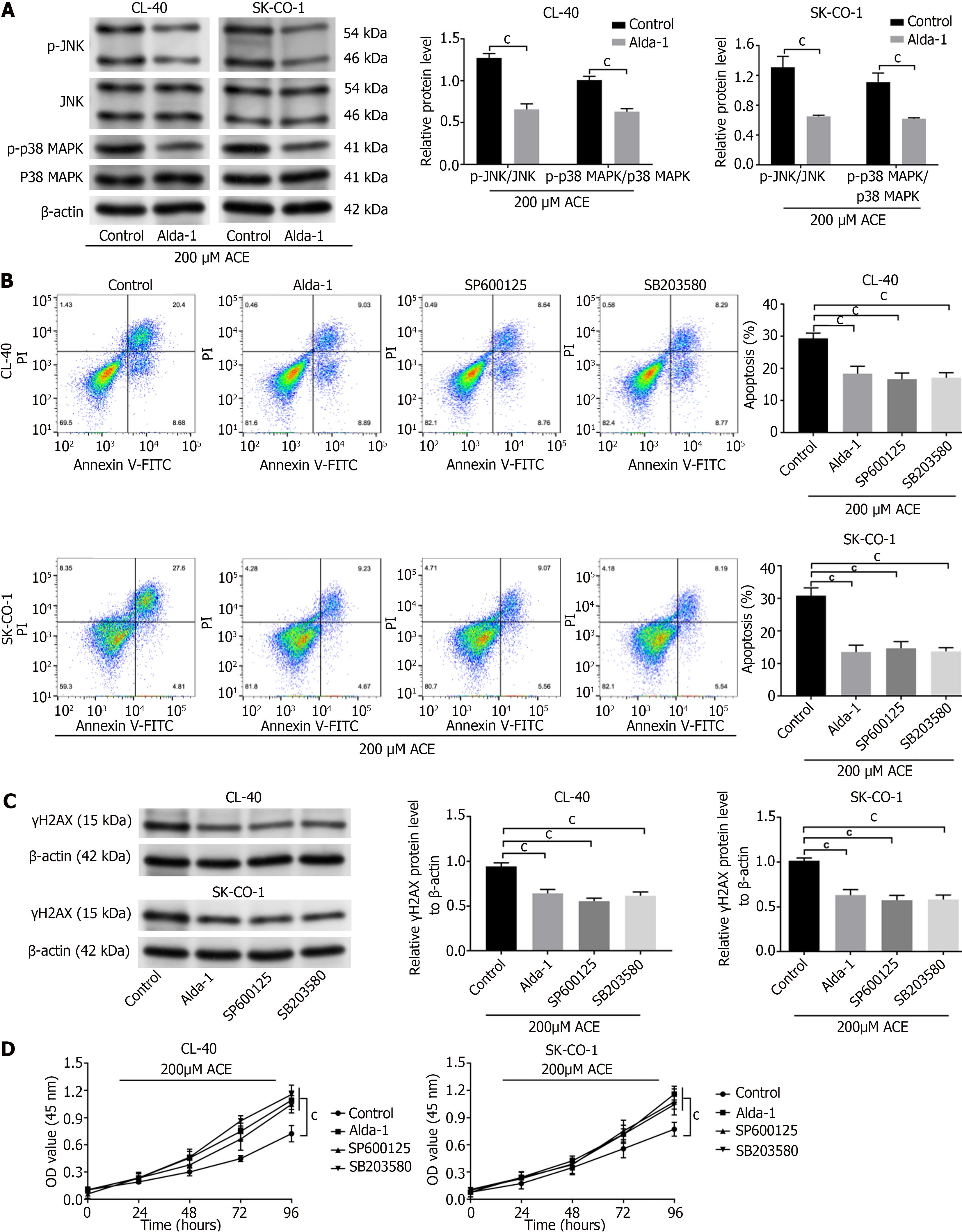
Figure 5 Aldehyde dehydrogenase 2 family member inhibited MAPK-apoptosis and DNA damage by regulating acetaldehyde in colorectal cancer cells.
A: Phosphorylation of JNK and P38 MAPK in CL-40 and SK-CO-1 cells in the presence of acetaldehyde (ACE) (200 μM) were measured by western blot; B: Flow cytometric analysis of apoptosis of CL-40 and SK-CO-1 cells in the presence of ACE; C: Detection of protein level of γH2AX in CL-40 and SK-CO-1 cells with the presence of ACE via western blot; D: The cell viability in CL-40 and SK-CO-1 cells with the presence of ACE was measured by cell counting kit-8. Data are displayed as the mean ± SD. cP < 0.001.
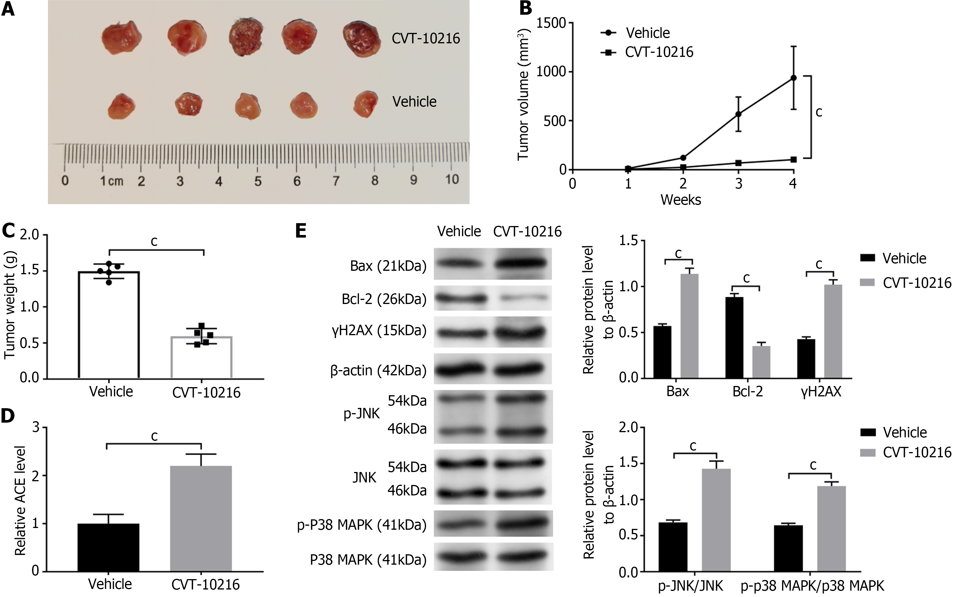
Figure 6 Aldehyde dehydrogenase 2 family member-deficiency leads to accumulated acetaldehyde and increased DNA damage in vivo.
A: The tumor growth in xenograft tumor mice model; B: The tumor volumes in shRNA aldehyde dehydrogenase 2 family member-SK-CO-1 mouse xenograft models treatment of imatinib; C: The mice were killed, and the tumor weight was assessed; D: Relative quantification of acetaldehyde of mice tumor tissues; E: Detection of Bax, Bcl-2, γH2AX, p-JNK/JNK, and p-P38 MAPK/P38 MAPK protein levels in tumor tissue by western blot. Results are the mean ± SD of triplicate samples. t-test, cP < 0.001.
- Citation: Yu M, Chen Q, Lu YP. Aldehyde dehydrogenase 2 family member repression promotes colorectal cancer progression by JNK/p38 MAPK pathways-mediated apoptosis and DNA damage. World J Gastrointest Oncol 2024; 16(7): 3230-3240
- URL: https://www.wjgnet.com/1948-5204/full/v16/i7/3230.htm
- DOI: https://dx.doi.org/10.4251/wjgo.v16.i7.3230


