Copyright
©The Author(s) 2024.
World J Gastrointest Oncol. Jul 15, 2024; 16(7): 3211-3229
Published online Jul 15, 2024. doi: 10.4251/wjgo.v16.i7.3211
Published online Jul 15, 2024. doi: 10.4251/wjgo.v16.i7.3211
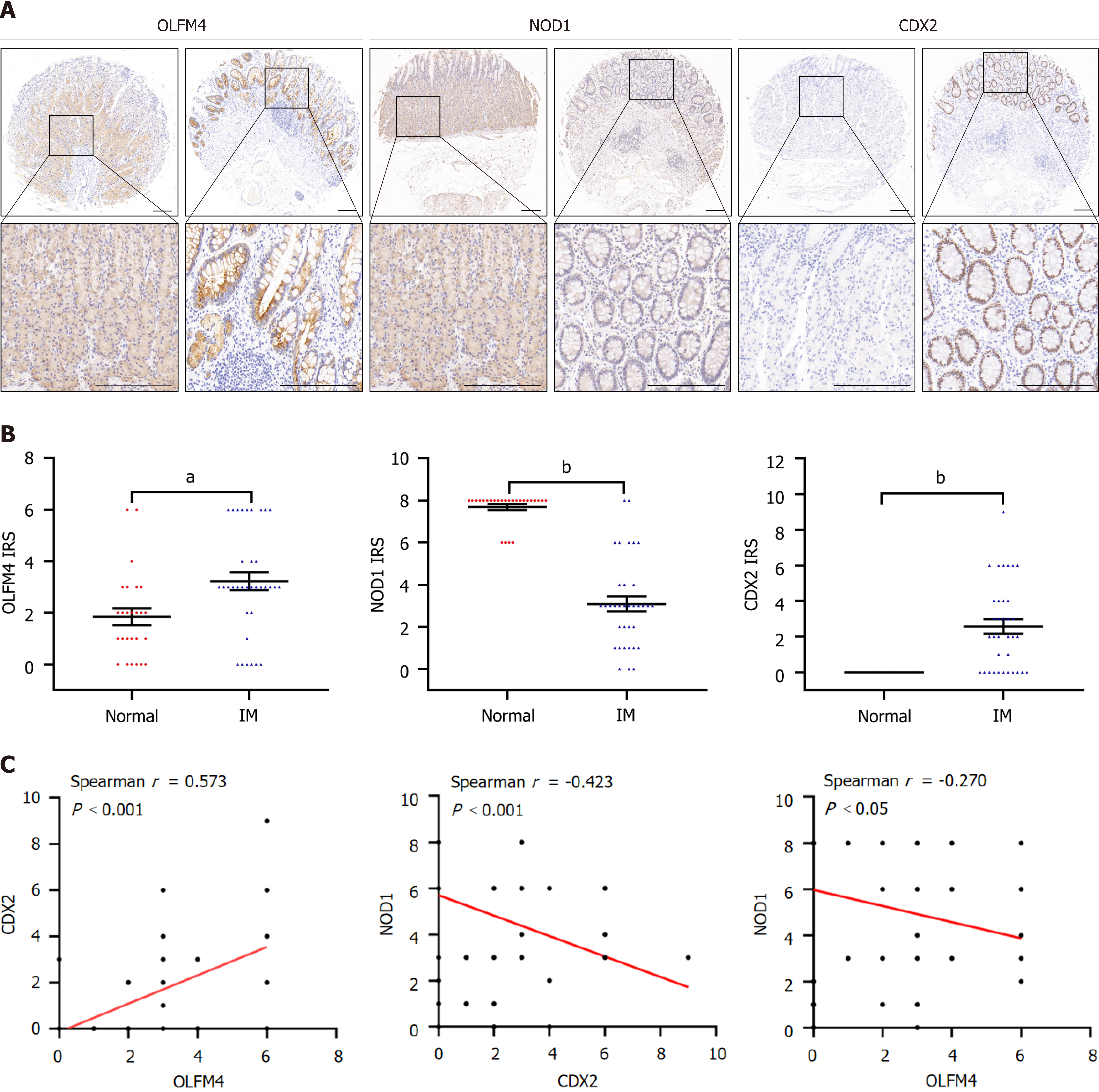
Figure 1 The olfactomedin 4/nucleotide-binding oligomerization domain 1/caudal-type homeobox gene 2 pathway is a characteristic of gastric intestinal metaplasia tissues.
A: The representative images of Immunohistochemistry (IHC) staining for olfactomedin 4 (OLFM4), nucleotide-binding oligomerization domain 1 (NOD1), and caudal-type homeobox gene 2 (CDX2) in normal and intestinal metaplasia (IM) tissues; B: OLFM4, NOD1, and CDX2 immunoreactivity scores in normal and IM tissues were compared; C: Correlation of OLFM4 with CDX2, NOD1 with CDX2, and OLFM4 with NOD1 in gastric tissues by Spearman’s correlation analysis. aP < 0.01, bP < 0.001 vs control group. OLFM4: Olfactomedin 4; NOD1: Nucleotide-binding oligomerization domain 1; CDX2: Caudal-type homeobox gene 2; IM: Intestinal metaplasia.
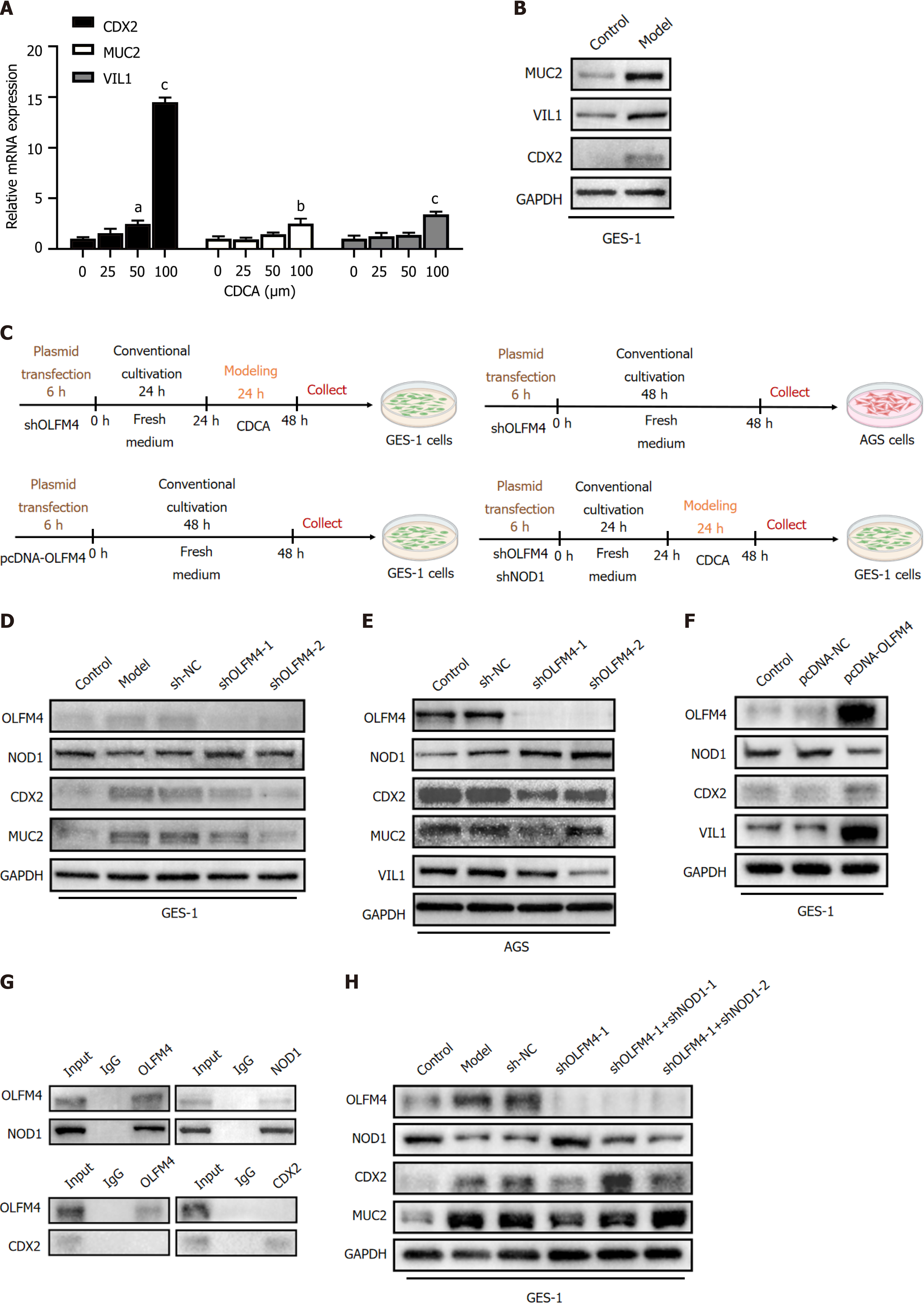
Figure 2 Olfactomedin 4 exhibited direct binding and subsequent down-regulation of nucleotide-binding oligomerization domain 1, thereby sustaining the activation of caudal-type homeobox gene 2 and promoting the progression of intestinal metaplasia.
A: Relative mRNA expressions of caudal-type homeobox gene 2 (CDX2), MUCIN 2 (MUC2), and villin 1 (VIL1) on stimulation of chenodeoxycholic acid (CDCA) in GES-1 cells (n = 3); B: Western blot (WB) detection of MUC2, VIL1, and CDX2 in GES-1 cells treated with CDCA (100 μM) for 24 h; C: Timeline of the cell experiments design for pathway regulatory verification; D: Short hairpin RNAs (shRNAs) targeting olfactomedin 4 (OLFM4) were transfected to GES-1 cells. Subsequently, the cells were treated with CDCA (100 μM) for 24 h. WB detection of OLFM4, nucleotide-binding oligomerization domain 1 (NOD1), CDX2, and MUC2; E: AGS cells were transfected with OLFM4 shRNAs. WB detection of OLFM4, NOD1, CDX2, MUC2, and VIL1; F: OLFM4-pcDNAs were transfected to GES-1 cells. WB detection of OLFM4, NOD1, CDX2, and VIL1; G: Co-immunoprecipitation detection of the interaction between OLFM4 and NOD1, and the interaction between OLFM4 and CDX2 in GES-1 cells; H: OLFM4 and NOD1 shRNAs were co-transfected to GES-1 cells. Subsequently, the cells were treated with CDCA (100 μM) for 24 h. WB detection of OLFM4, NOD1, CDX2, and MUC2. aP < 0.05, bP < 0.01, cP < 0.001 vs control group. OLFM4: Olfactomedin 4; NOD1: Nucleotide-binding oligomerization domain 1; CDX2: Caudal-type homeobox gene 2; MUC2: MUCIN 2; VIL1: Villin 1; CDCA: Chenodeoxycholic acid.
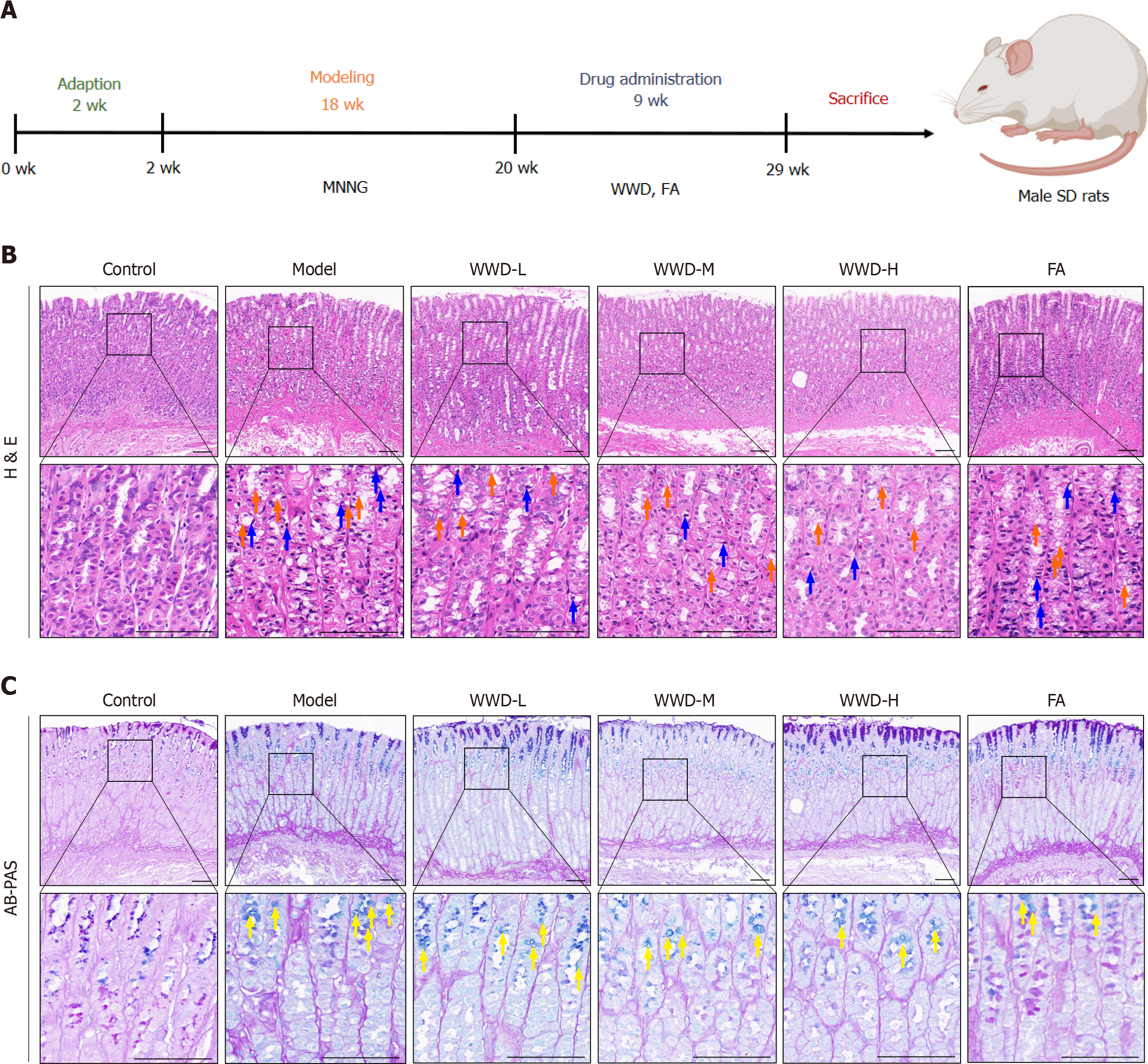
Figure 3 Weiwei Decoction improves gastric mucosal histological lesions.
A: Timeline of the animal experiments design for Weiwei Decoction treatment; B: Representative hematoxylin and eosin-stained images of the gastric mucosa in each group (n = 3). Vacuolation changes (orange arrow), pyknotic and hyperchromatic nuclei (blue arrow); C: Representative Alcian blue-Periodic acid-Schiff-stained images of the gastric mucosa in each group (n = 3). Blue-stained cavities (yellow arrow). Scale bar: 100 μm. WWD: Weiwei Decoction; FA: Folic acid; HE: Hematoxylin and eosin; AB-PAS: Alcian blue-Periodic acid-Schiff.
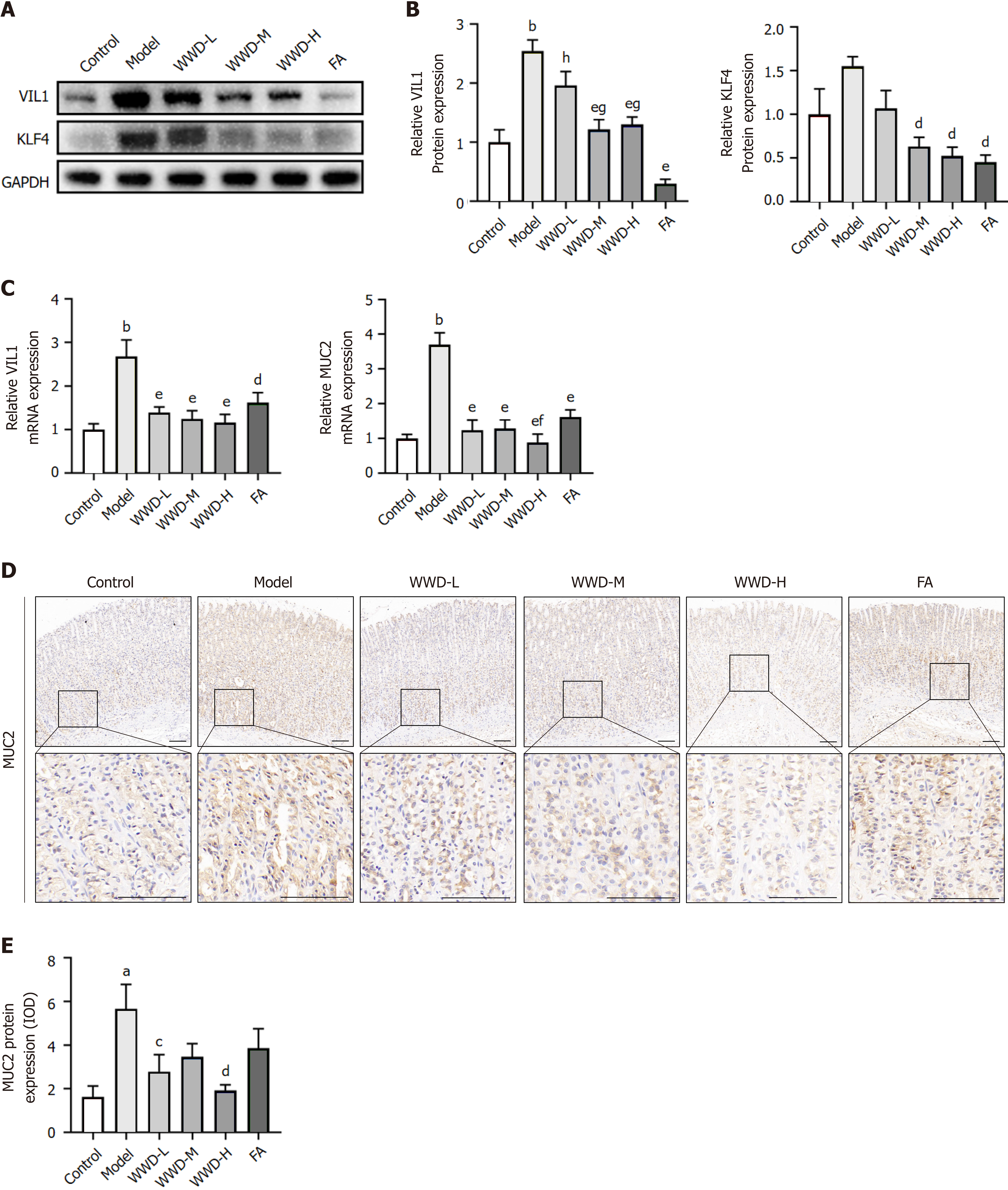
Figure 4 Weiwei Decoction reduces the expression of intestinal markers KLF transcription factor 4, villin 1, and MUCIN 2 in intestinal metaplasia rats.
A: Western blot detection of villin 1 (VIL1) and KLF transcription factor 4 (KLF4) in rat gastric mucosa; B: Relative protein expression of VIL1 and KLF4 (n = 4); C: Relative mRNA expression of MUCIN 2 (MUC2) and VIL1 in gastric mucosa (n = 5); D: Immunohistochemistry detection of MUC2, representative images of the gastric mucosa in each group. Scale bar: 100 μm; E: Quantitative analysis of MUC2 protein expression in gastric tissues (n = 3). aP < 0.01, bP < 0.001 vs control group; cP < 0.05, dP < 0.01, eP < 0.001 vs model group; fP < 0.05, gP < 0.01, hP < 0.001 vs folic acid group. VIL1: Villin 1; KLF4: KLF transcription factor 4; WWD: Weiwei Decoction; FA: Folic acid; MUC2: MUCIN 2.
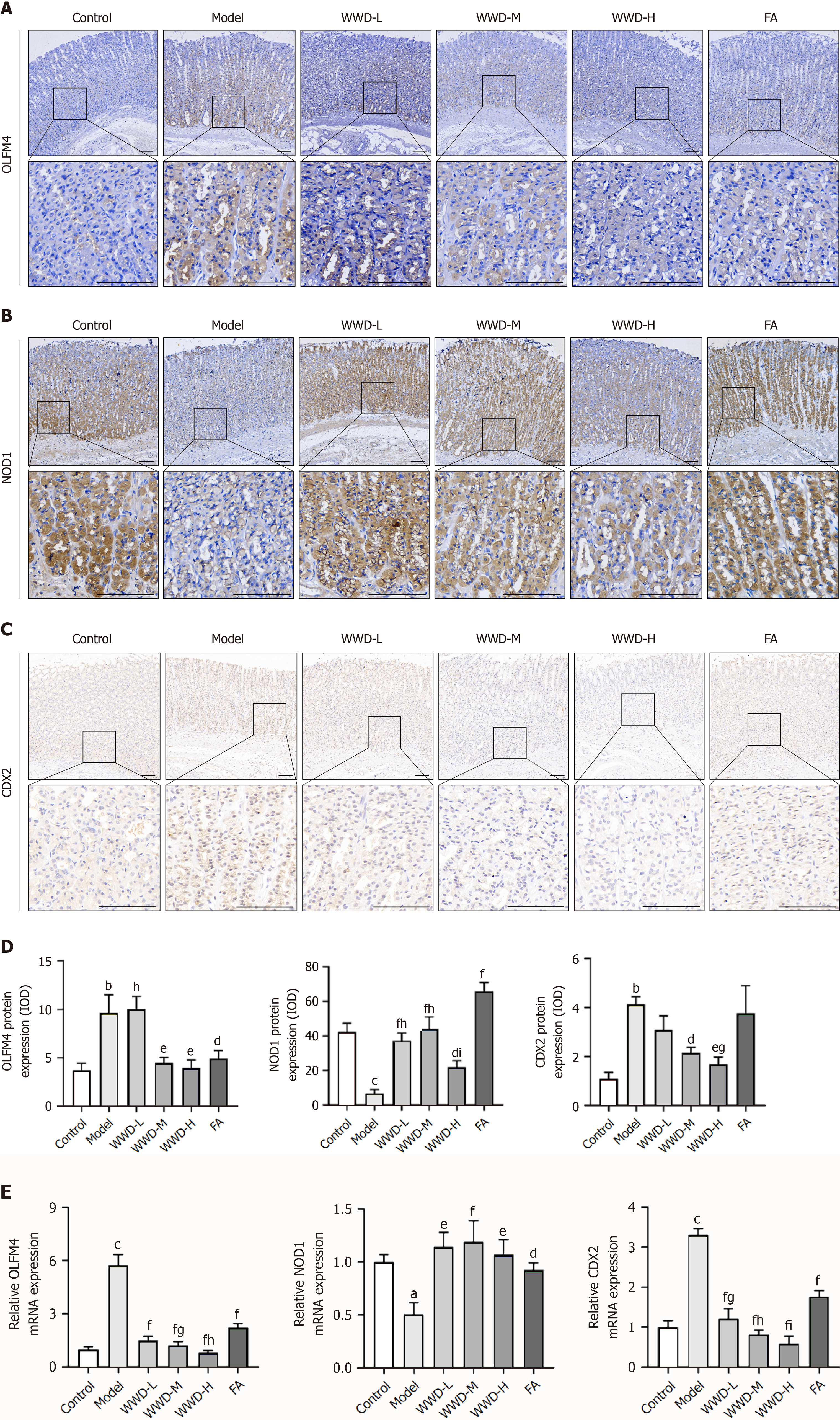
Figure 5 Weiwei Decoction suppresses olfactomedin 4 expression and restores nucleotide-binding oligomerization domain 1 level, consequently reducing caudal-type homeobox gene 2 in intestinal metaplasia rats.
A-C: Immunohistochemistry detection of olfactomedin 4 (OLFM4), nucleotide-binding oligomerization domain 1 (NOD1), and caudal-type homeobox gene 2 (CDX2), representative images of gastric mucosa in each group. Scale bar: 100 μm; D: Quantitative analysis of OLFM4, NOD1, and CDX2 protein expression in gastric mucosa (n = 3); E: Relative mRNA expression of OLFM4, NOD1, and CDX2 in gastric mucosa (n = 5). aP < 0.05, bP < 0.01, cP < 0.001 vs control group; dP < 0.05, eP < 0.01, fP < 0.001 vs model group; gP < 0.05, hP < 0.01, iP < 0.001 vs folic acid group. OLFM4: Olfactomedin 4; NOD1: Nucleotide-binding oligomerization domain 1; CDX2: Caudal-type homeobox gene 2; WWD: Weiwei Decoction; FA: Folic acid.
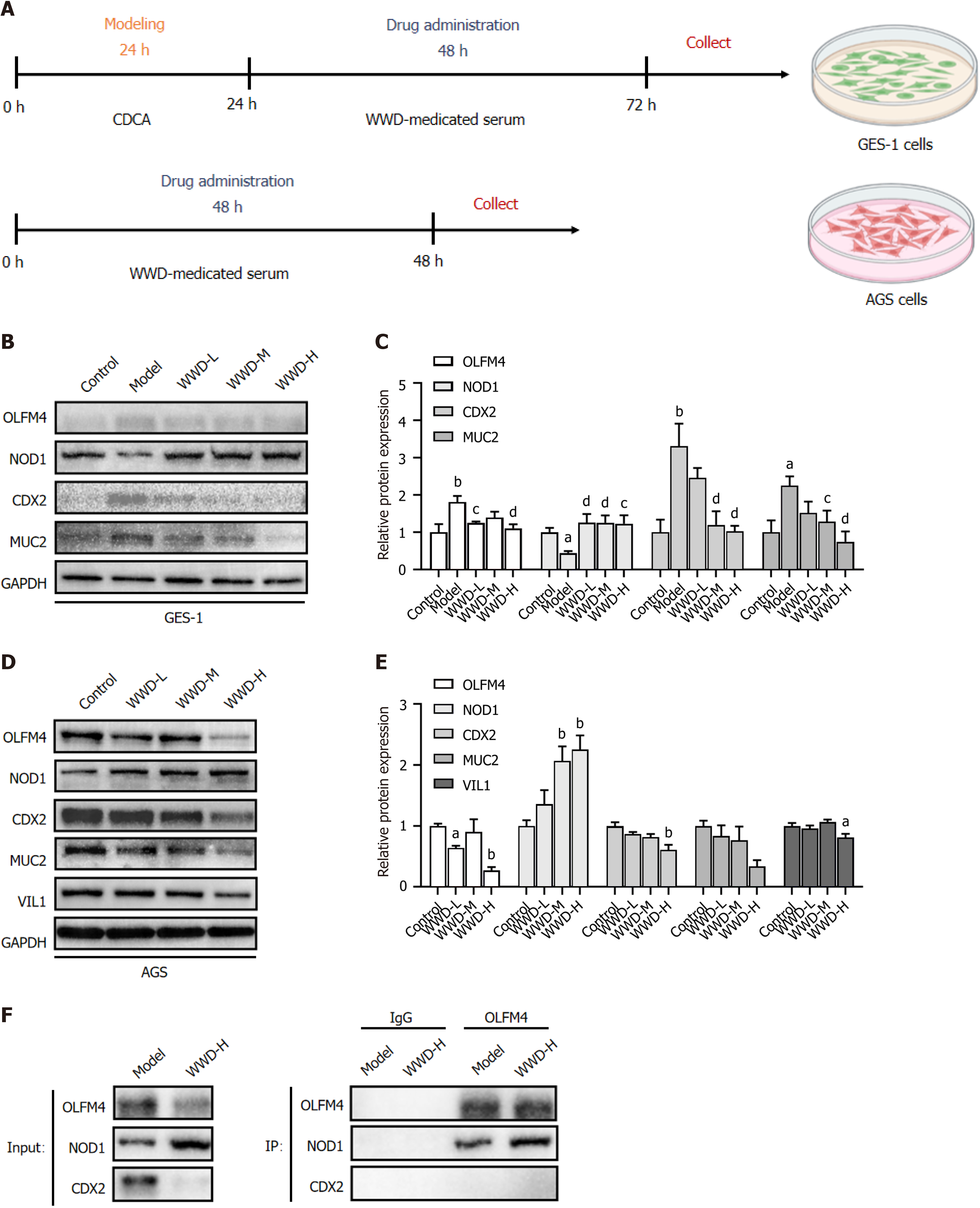
Figure 6 Weiwei Decoction-medicated serum attenuates olfactomedin 4 level and simultaneously strengthens olfactomedin 4 and nucleotide-binding oligomerization domain 1 binding, thereby enhancing nucleotide-binding oligomerization domain 1 expression and consequently mitigating caudal-type homeobox gene 2 in gastric cells.
A: Timeline of the cell experiments design for Weiwei Decoction (WWD)-medicated serum treatment; B: GES-1 cells were treated with chenodeoxycholic acid (CDCA) for 24 h and then disposed to 10% (V/V) of the corresponding WWD-medicated serum for 48 h. WB detection of olfactomedin 4 (OLFM4), nucleotide-binding oligomerization domain 1 (NOD1), caudal-type homeobox gene 2 (CDX2), and MUCIN 2 (MUC2); C: Relative protein expression of OLFM4, NOD1, CDX2, and MUC2 in GES-1 cells (n = 3); D: AGS cells were disposed to 10% (V/V) of the corresponding WWD-medicated serum for 48 h. WB detection of OLFM4, NOD1, CDX2, MUC2, and villin 1 (VIL1); E: Relative protein expression of OLFM4, NOD1, CDX2, MUC2, and VIL1 in AGS cells (n = 3); F: GES-1 cells were treated with CDCA for 24 h and then disposed to 10% (V/V) of the WWD-H medicated serum for 48 h. Co-immunoprecipitation detection of the interaction between OLFM4 and NOD1, and the interaction between OLFM4 and CDX2 in GES-1 cells. aP < 0.05, bP < 0.01 vs control group; cP < 0.05, dP < 0.01 vs model group. OLFM4: Olfactomedin 4; NOD1: Nucleotide-binding oligomerization domain 1; CDX2: Caudal-type homeobox gene 2; WWD: Weiwei Decoction; MUC2: MUCIN 2; VIL1: Villin 1.
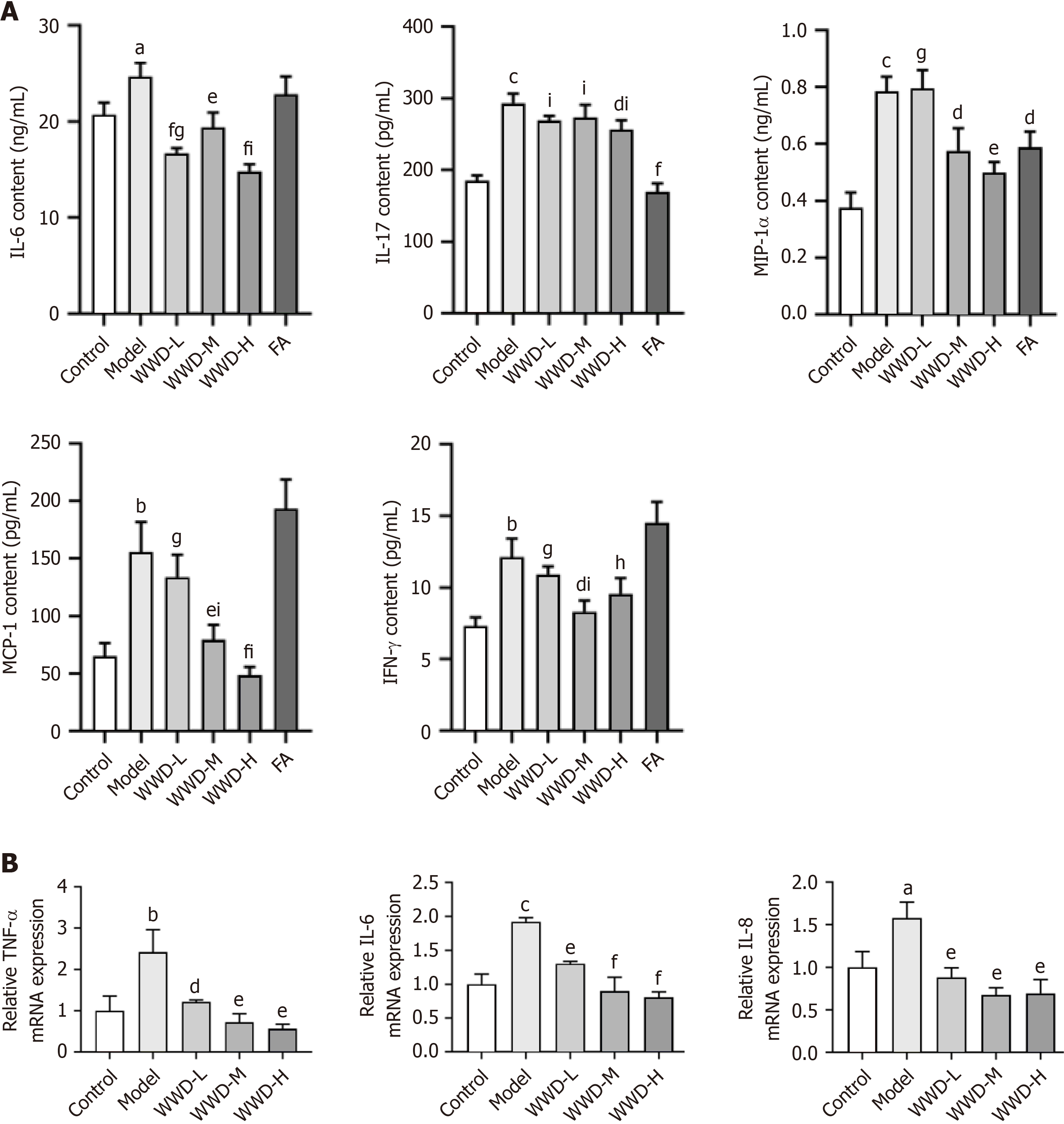
Figure 7 Weiwei Decoction inhibits cytokines and chemokines in gastric intestinal metaplasia.
A: Enzyme-linked immunosorbent assay was utilized to detect interferon-gamma, interleukin (IL)-6, IL-17, macrophage chemoattractant protein-1, and macrophage inflammatory protein 1 alpha in intestinal metaplasia rats serum (n = 6); B: Relative mRNA expression of tumor necrosis factor alpha, IL-6, and IL-8 in GES-1 cells (n = 3). aP < 0.05, bP < 0.01, cP < 0.001 vs control group; dP < 0.05, eP < 0.01, fP < 0.001 vs model group; gP < 0.05, hP < 0.01, iP < 0.001 vs folic acid group. IL: Interleukin; MIP: Macrophage inflammatory protein; MCP: Macrophage chemoattractant protein; IFN: Interferon; WWD: Weiwei Decoction; FA: Folic acid; TNF: Tumor necrosis factor.
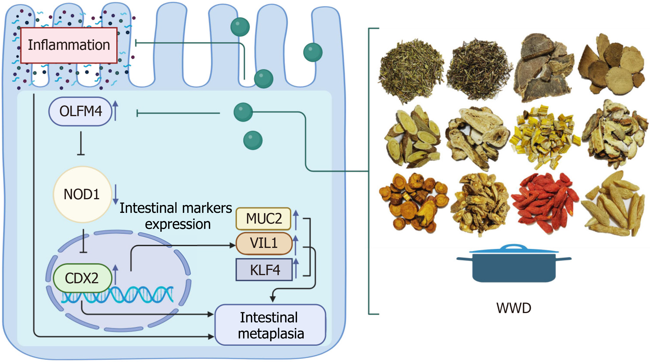
Figure 8 A schematic model of the olfactomedin 4/nucleotide-binding oligomerization domain 1/caudal-type homeobox gene 2 pathway in gastric intestinal metaplasia cells and the mechanism of Weiwei Decoction in the treatment of gastric intestinal metaplasia through this pathway.
OLFM4: Olfactomedin 4; NOD1: Nucleotide-binding oligomerization domain 1; CDX2: Caudal-type homeobox gene 2; VIL1: Villin 1; KLF4: KLF transcription factor 4; WWD: Weiwei Decoction; MUC2: MUCIN 2.
- Citation: Zhou DS, Zhang WJ, Song SY, Hong XX, Yang WQ, Li JJ, Xu JQ, Kang JY, Cai TT, Xu YF, Guo SJ, Pan HF, Li HW. Weiwei Decoction alleviates gastric intestinal metaplasia through the olfactomedin 4/nucleotide-binding oligomerization domain 1/caudal-type homeobox gene 2 signaling pathway. World J Gastrointest Oncol 2024; 16(7): 3211-3229
- URL: https://www.wjgnet.com/1948-5204/full/v16/i7/3211.htm
- DOI: https://dx.doi.org/10.4251/wjgo.v16.i7.3211


