Copyright
©The Author(s) 2024.
World J Gastrointest Oncol. Jun 15, 2024; 16(6): 2663-2672
Published online Jun 15, 2024. doi: 10.4251/wjgo.v16.i6.2663
Published online Jun 15, 2024. doi: 10.4251/wjgo.v16.i6.2663
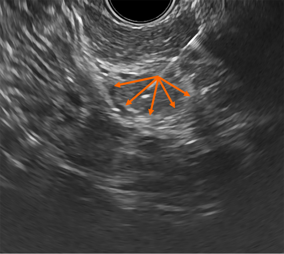
Figure 1 Ultrasound image of the pancreatic body including a hypoechoic lesion (in the center) with clear boundaries.
Orange arrows indicate the directions of the needle movements during fine-needle aspiration using the “fanning” technique.
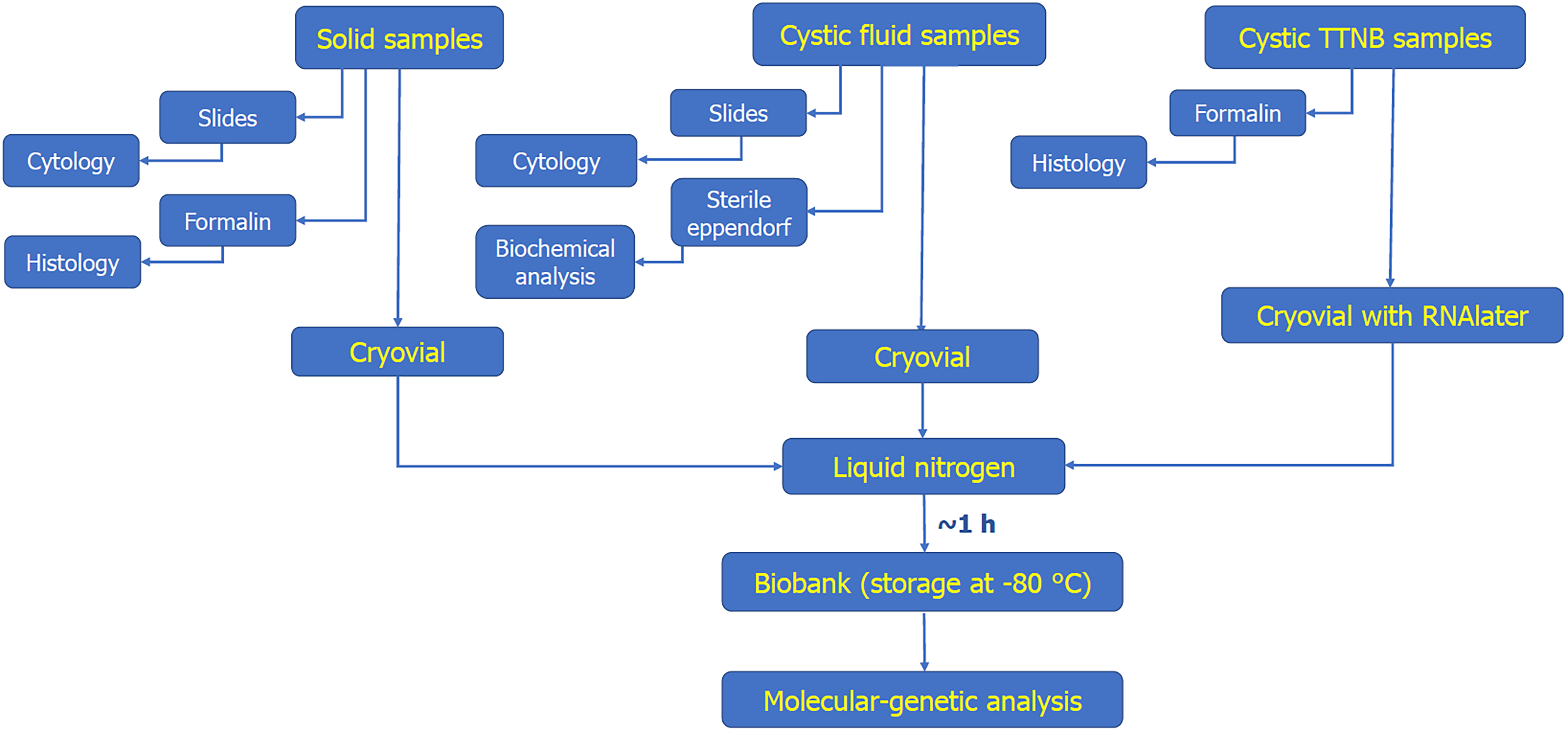
Figure 2 Strategy for processing biopsy samples obtained from fine-needle aspiration under endoscopic ultrasound guidance.
The steps for processing biopsy samples for biobanking are highlighted in yellow. TTNB: Through-the-needle biopsy.
- Citation: Seyfedinova SS, Freylikhman OA, Sokolnikova PS, Samochernykh KA, Kostareva AA, Kalinina OV, Solonitsyn EG. Fine-needle aspiration technique under endoscopic ultrasound guidance: A technical approach for RNA profiling of pancreatic neoplasms. World J Gastrointest Oncol 2024; 16(6): 2663-2672
- URL: https://www.wjgnet.com/1948-5204/full/v16/i6/2663.htm
- DOI: https://dx.doi.org/10.4251/wjgo.v16.i6.2663


