Copyright
©The Author(s) 2024.
World J Gastrointest Oncol. May 15, 2024; 16(5): 1773-1786
Published online May 15, 2024. doi: 10.4251/wjgo.v16.i5.1773
Published online May 15, 2024. doi: 10.4251/wjgo.v16.i5.1773
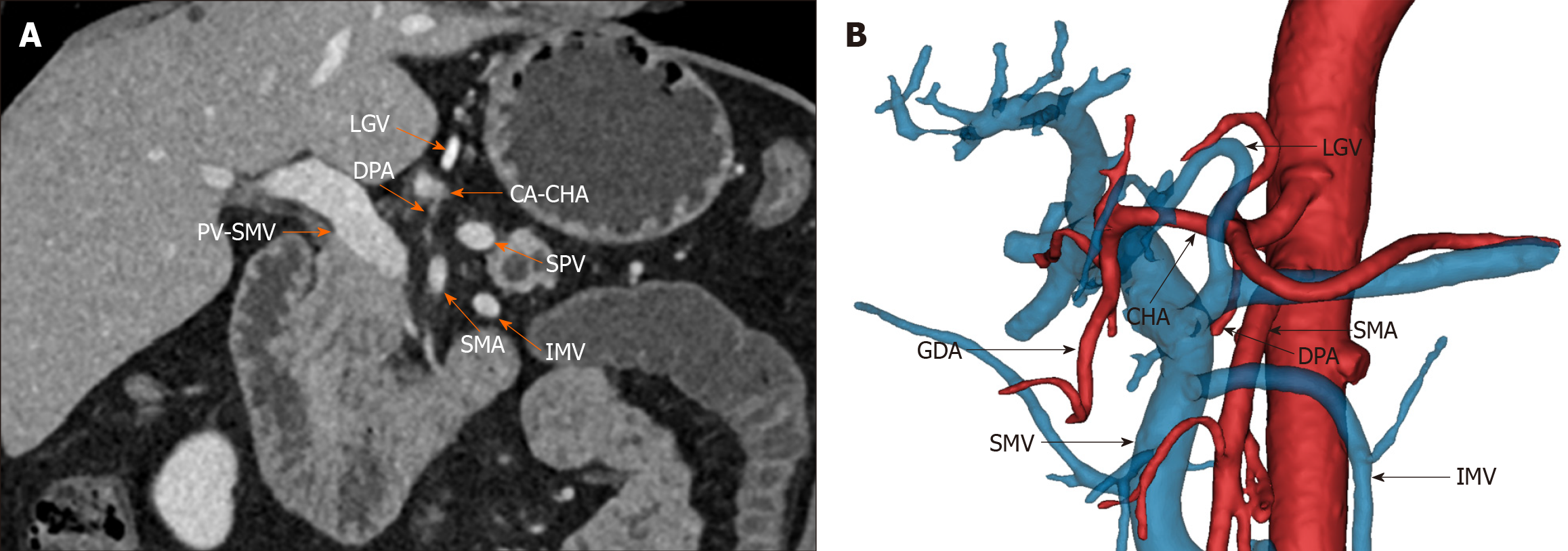
Figure 1 The preoperative imaging of Heidelberg triangle.
A: Preoperative contrast-enhanced computed tomography image of the abdomen. The orange arrowhead points to vessels around the Heidelberg triangle. The portal vein-superior mesenteric vein, celiac axis-common hepatic artery (CHA), and superior mesenteric artery represent the three boundary vessels of the Heidelberg triangle. The dorsal pancreatic artery (DPA) in the figure originates from the CHA and passes through the triangle, reflecting one of the numerous pathways of DPA. The left gastric vein (LGV), splenic vein (SPV), and inferior mesenteric vein (IMV) typically pass anteriorly to the triangle; B: Frontal view of the Heidelberg triangle generated by three-dimensional reconstruction. The black arrowhead points to vessels around the Heidelberg triangle. The image reveals the DPA coursing within the triangle and the LGV, SPV, and IMV traversing anteriorly to the triangle. GDA: Gastroduodenal artery; PV: Portal vein; SMV: Superior mesenteric vein; CA: Celiac axis; CHA: Common hepatic artery; SMA: Superior mesenteric artery; DPA: Dorsal pancreatic artery; LGV: Left gastric vein; SPV: Splenic vein; IMV: Inferior mesenteric vein.
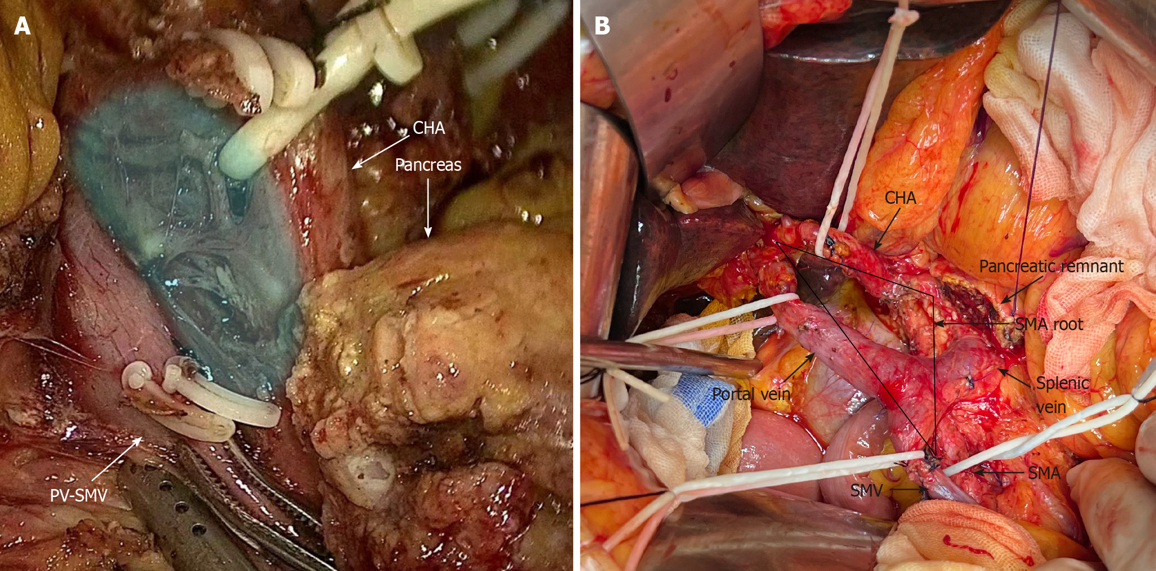
Figure 2 Intraoperative image of the Heidelberg triangle dissection.
A: Intraoperative image before Heidelberg triangle dissection. At this stage, the superior mesenteric artery (SMA) is located on the dorsal side of the pancreatic remnant, and the portal vein (PV)-superior mesenteric vein (SMV), common hepatic artery (CHA), and pancreas are visible. The blue shading illustrates the boundaries of the Heidelberg triangle, encompassing vascular, lymphatic, and neural fiber tissues within the triangle; B: Intraoperative image after Heidelberg triangle dissection. The black arrowhead points to vessels and tissues around the Heidelberg triangle. The black triangle shows the range of Heidelberg triangle dissection. After thorough clearance, the precise boundaries of the triangle and its three bordering vessels, namely, the PV-SMV, CHA, and SMA, are clearly visible. SMV: Superior mesenteric vein; SMA: Superior mesenteric artery; CHA: Common hepatic artery; PV: Portal vein.
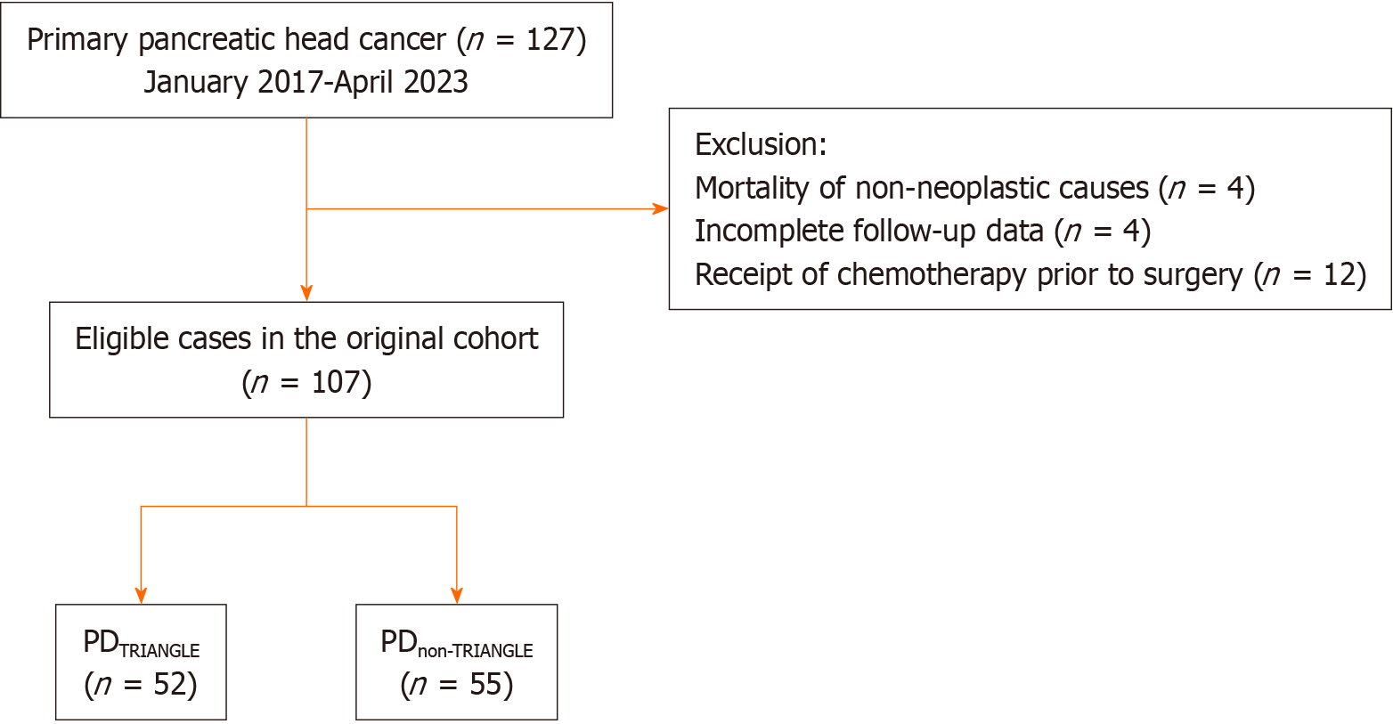
Figure 3 Flow diagram of the study cohort.
Pancreaticoduodenectomy (PD)TRIANGLE, patients underwent standard lymphadenectomy along with Heidelberg triangle dissection during pancreaticoduodenectomy; PDnon-TRIANGLE, patients underwent standard lymphadenectomy during pancreaticoduodenectomy without Heidelberg triangle dissection. PD: Pancreaticoduodenectomy.
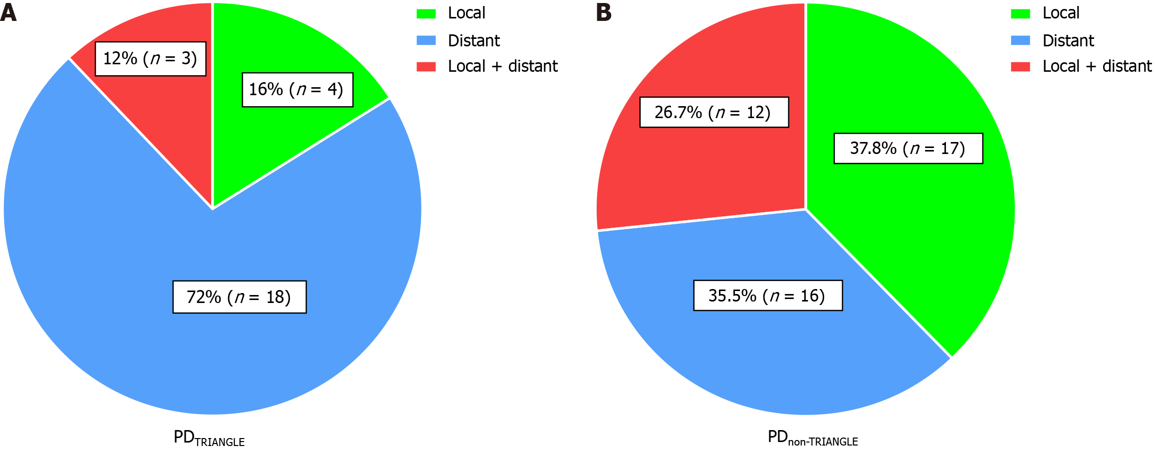
Figure 4 The recurrence patterns in both groups.
A: Recurrence pattern in the Pancreaticoduodenectomy (PD)TRIANGLE group. The proportions of local, distant, and combined local and distant recurrences were 16%, 72%, and 12%, respectively; B: Recurrence pattern in the PDnon-TRIANGLE group. The percentages of local, distant, and combined local and distant recurrences were 37.8%, 35.5%, and 26.7%, respectively. PD: Pancreaticoduodenectomy.
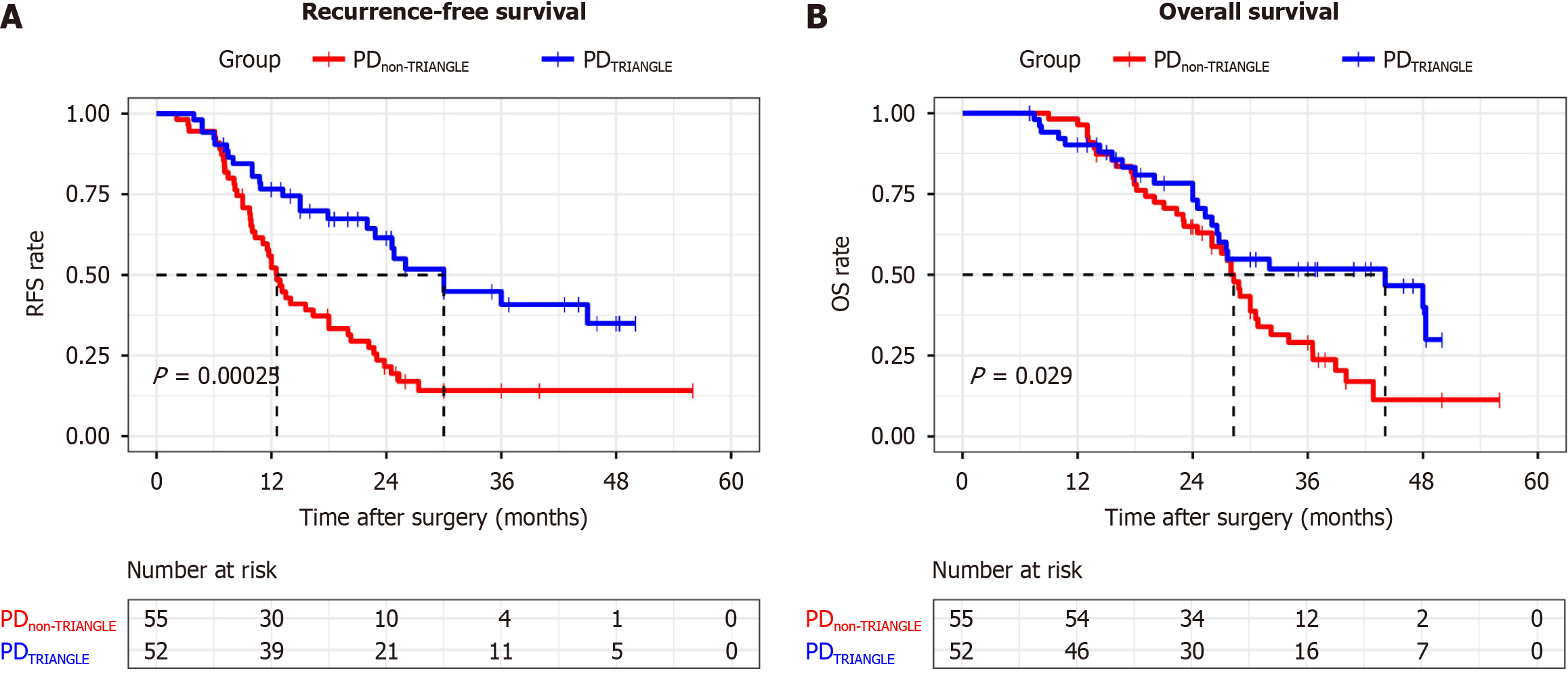
Figure 5 Kaplan-Meier curves of both groups.
A: Kaplan-Meier curve of recurrence-free survival (RFS) between the Pancreaticoduodenectomy (PD)TRIANGLE and PDnon-TRIANGLE groups. The median RFS was notably longer in the PDTRIANGLE group at 30.0 months than in the PDnon-TRIANGLE group at 12.6 months (P < 0.001); B: Kaplan-Meier curve of overall survival (OS) between the PDTRIANGLE and PDnon-TRIANGLE groups. The median OS was significantly better in the PDTRIANGLE group than in the PDnon-TRIANGLE group (44.1 months vs 28.3 months, P = 0.029). PD: Pancreaticoduodenectomy; RFS: Recurrence-free survival.
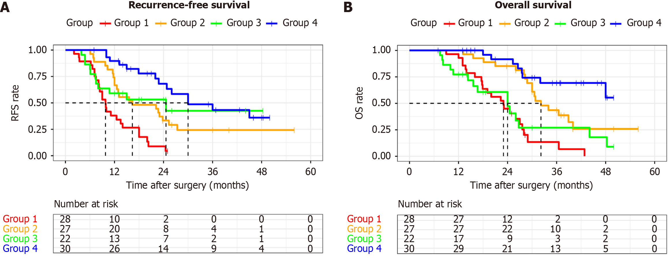
Figure 6 Kaplan-Meier curves of the four groups.
A: Kaplan-Meier curve of recurrence-free survival (RFS) between the four groups. The median RFS (mRFS) for groups 1, 2, 3, and 4 were 9.8, 16.4, 24.6, and 30.0 months, respectively. From group 1 to group 4, the mRFS progressively increased. The mRFS of group 1 was significantly shorter than that of groups 2, 3, and 4; group 4 exhibited a significantly longer mRFS than group 2 did; B: Kaplan-Meier curve of overall survival (OS) between the four groups. The median OS (mOS) for groups 1, 2, 3, and 4 were 23.0, 32.2, 24.0 months, and not reached, respectively. The mOS of groups 2 and 4 was significantly longer than that of groups 1 and 3, and the mOS of group 4 was significantly longer than that of group 2. Group 1: No clearance of the Heidelberg triangle and not receiving ≥ 6 months of adjuvant chemotherapy; Group 2: No clearance of the Heidelberg triangle but receiving ≥ 6 months of adjuvant chemotherapy; Group 3: Clearance of the Heidelberg triangle but not receiving ≥ 6 months of adjuvant chemotherapy; Group 4: Clearance of the Heidelberg triangle and receiving ≥ 6 months of adjuvant chemotherapy. RFS: Recurrence-free survival; OS: Overall survival.
- Citation: Chen JH, Zhu LY, Cai ZW, Hu X, Ahmed AA, Ge JQ, Tang XY, Li CJ, Pu YL, Jiang CY. TRIANGLE operation, combined with adequate adjuvant chemotherapy, can improve the prognosis of pancreatic head cancer: A retrospective study. World J Gastrointest Oncol 2024; 16(5): 1773-1786
- URL: https://www.wjgnet.com/1948-5204/full/v16/i5/1773.htm
- DOI: https://dx.doi.org/10.4251/wjgo.v16.i5.1773


