Copyright
©The Author(s) 2024.
World J Gastrointest Oncol. Apr 15, 2024; 16(4): 1192-1203
Published online Apr 15, 2024. doi: 10.4251/wjgo.v16.i4.1192
Published online Apr 15, 2024. doi: 10.4251/wjgo.v16.i4.1192
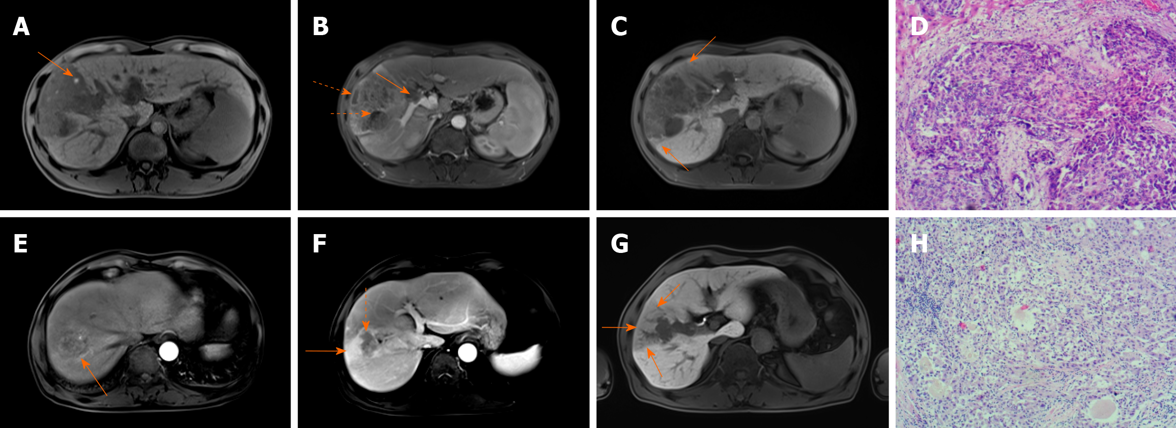
Figure 1 Typical cases of early recurrence of hepatocellular carcinoma after resection.
A-D: A 45-year-old male with pathologically proven hepatocellular carcinoma (HCC) [China liver cancer (CNLC) stage IIIa] suffering from recurrence 3 months after surgery. Axial T1-weighted imaging showed hyperintensity signal in the tumor indicating intratumoral hemorrhage (A, arrow). Gadolinium ethoxybenzyl diethylenetriamine pentaacetic acid (Gd-EOB-DTPA)-enhanced magnetic resonance imaging (MRI) showed large vessel invasion with filling defect in the portal vein (b, arrow) and non-enhanced necrotic area (B, dashed arrow). The tumor showed irregular or nodular like shape with incomplete capsule and peritumoral hypointensity (C, arrows) in the hepatobiliary phase. The pathological result was moderately differentiated HCC with microvascular invasion (D); E-H: A 60-year-old male with moderately differentiated HCC experienced recurrence 6 months after surgery (CNLC stage IIIa). Gd-EOB-DTPA-enhanced MRI showed peritumoral enhancement manifesting patchy enhancing areas around the tumor in the arterial and venous phase (E and F, arrows). The tumor was inhomogeneously nonrim arterial phase hyperenhancement and nonperipheral washout in the portal venous phase with non-enhanced necrotic area (G, arrow). Peritumoral hypointensity was seen in the hepatobiliary phase (F, arrows). The pathological result was moderately differentiated HCC with microvascular invasion (H).
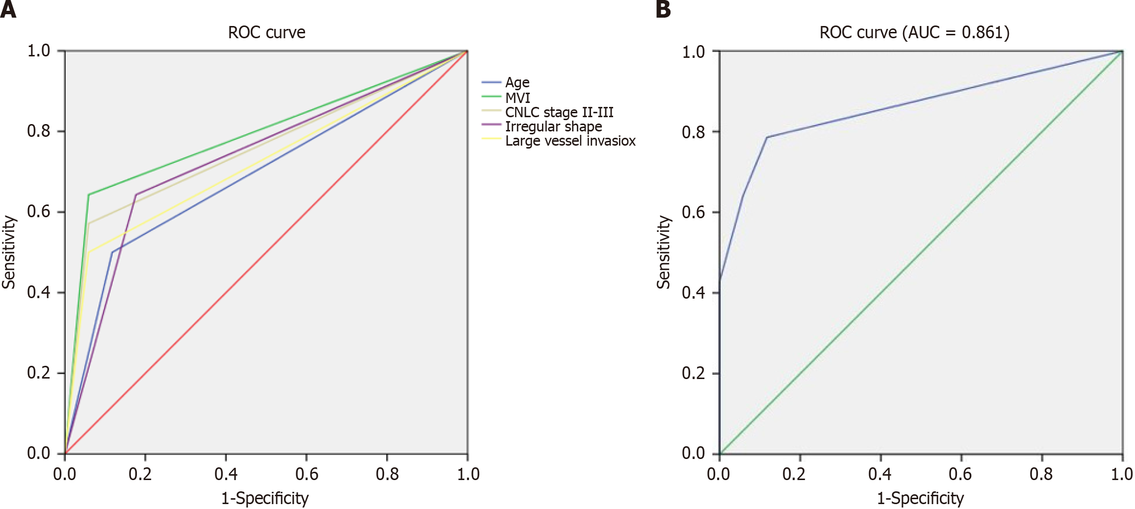
Figure 2 Receiver operating characteristics curves.
A and B: The receiver operating characteristics curves of the independent predictors for early post-operative hepatocellular carcinoma recurrence (A) and combining of multiple predictors (B). ROC: Receiver operating characteristics.
- Citation: Chen JP, Yang RH, Zhang TH, Liao LA, Guan YT, Dai HY. Pre-operative enhanced magnetic resonance imaging combined with clinical features predict early recurrence of hepatocellular carcinoma after radical resection. World J Gastrointest Oncol 2024; 16(4): 1192-1203
- URL: https://www.wjgnet.com/1948-5204/full/v16/i4/1192.htm
- DOI: https://dx.doi.org/10.4251/wjgo.v16.i4.1192


