Copyright
©The Author(s) 2024.
World J Gastrointest Oncol. Jan 15, 2024; 16(1): 244-250
Published online Jan 15, 2024. doi: 10.4251/wjgo.v16.i1.244
Published online Jan 15, 2024. doi: 10.4251/wjgo.v16.i1.244
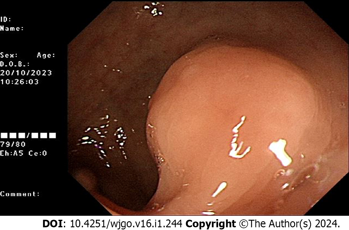
Figure 1 Colonoscopy reveals a submucosal protrusion located 15 cm from the anus.
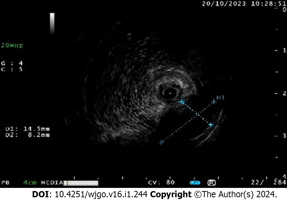
Figure 2 Ultrasound endoscopy displays a hypoechoic mass at the lesion, which is located in the lamina propria, with heterogeneous echogenicity, about 1.
0 cm in size.
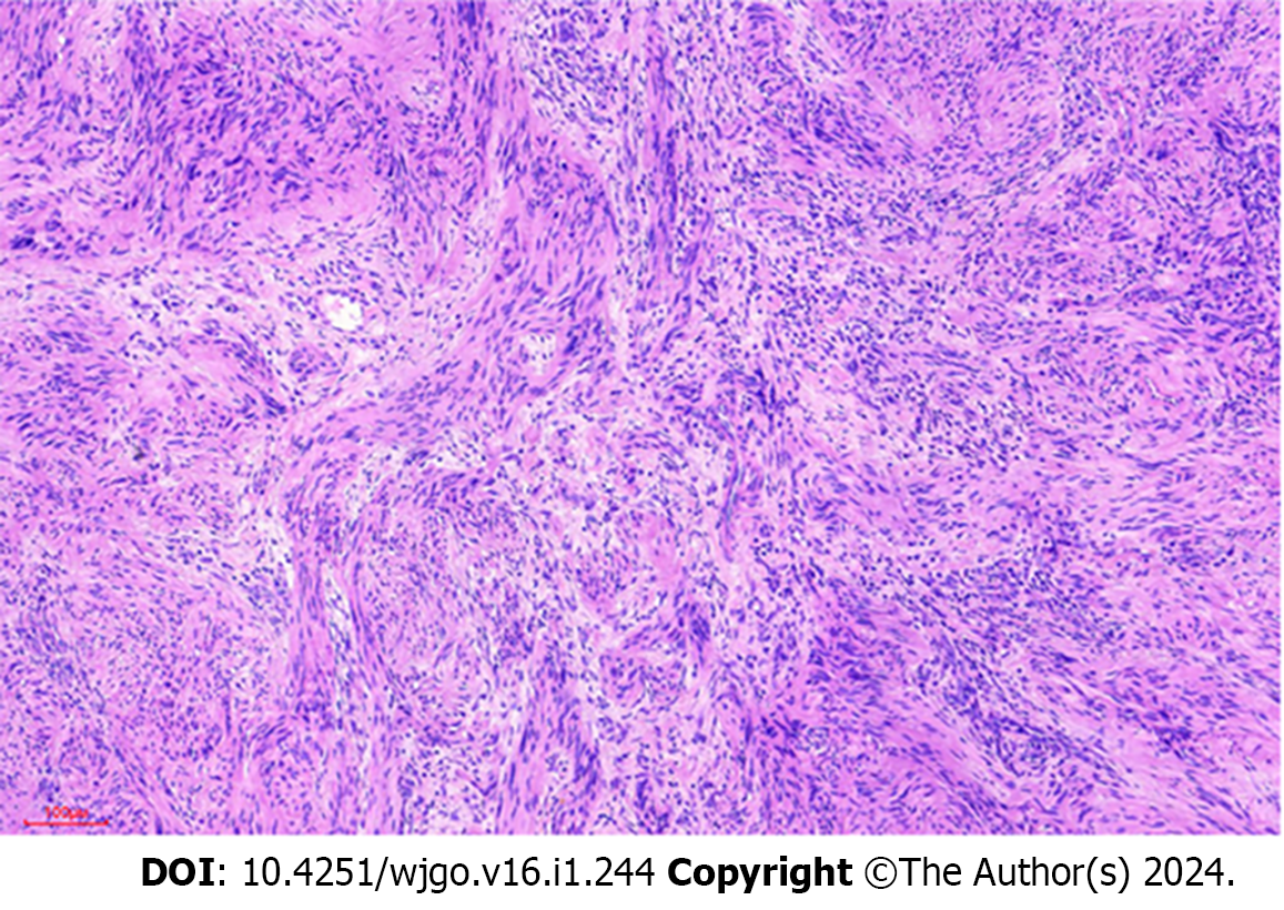
Figure 3 The Hematoxylin and eosin staining demonstrates the presence of spindle-shaped anisotropic cells exhibiting an intact peritoneum and the absence of extraperitoneal invasion.
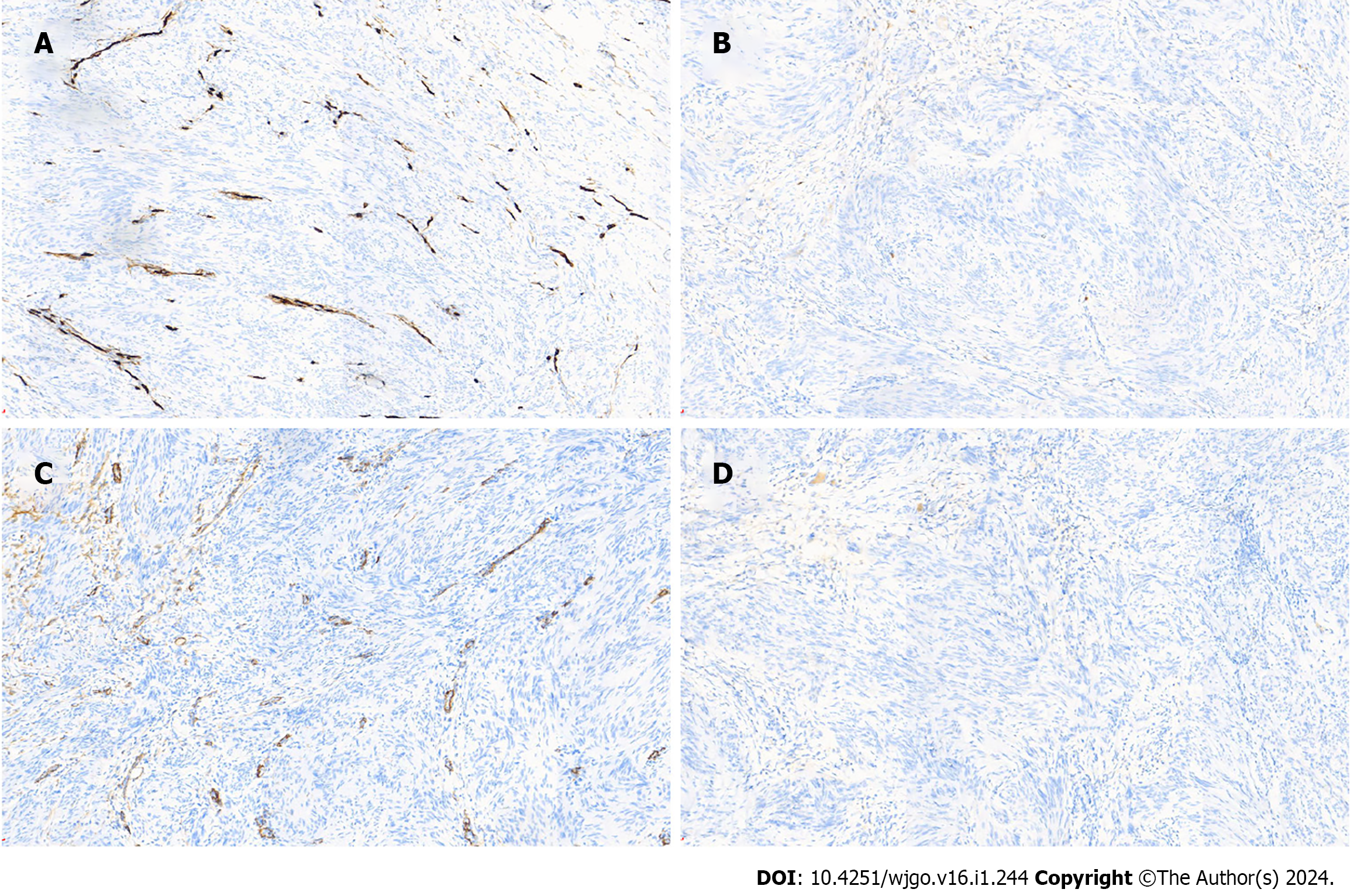
Figure 4 CD34, CD117, SMA, Dog-1 are negative in tumor cells.
A: CD34 is negative in tumor cells, while normal vascular structures are stained; B: CD117 is negative in tumor cells; C: SMA is negative in tumor cells; D: Dog-1 is negative in tumor cells.
Figure 5 SOX10 and S100.
A: S-100 is diffusely strongly positive in tumor cells; B: SOX-10 is strongly positive in tumor cells. SOX10 and S100 are positive. The immunohistochemical staining exhibits a consistent dark brown color.
Figure 6 Tumor.
A: Tumor of about 1.0 cm in size ; B: Tumor is observed as unencapsulated yellowish material.
- Citation: Li JY, Gao XZ, Zhang J, Meng XZ, Cao YX, Zhao K. Comprehensive evaluation of rare case: From diagnosis to treatment of a sigmoid Schwannoma: A case report. World J Gastrointest Oncol 2024; 16(1): 244-250
- URL: https://www.wjgnet.com/1948-5204/full/v16/i1/244.htm
- DOI: https://dx.doi.org/10.4251/wjgo.v16.i1.244


