Copyright
©The Author(s) 2022.
World J Gastrointest Oncol. Sep 15, 2022; 14(9): 1727-1738
Published online Sep 15, 2022. doi: 10.4251/wjgo.v14.i9.1727
Published online Sep 15, 2022. doi: 10.4251/wjgo.v14.i9.1727
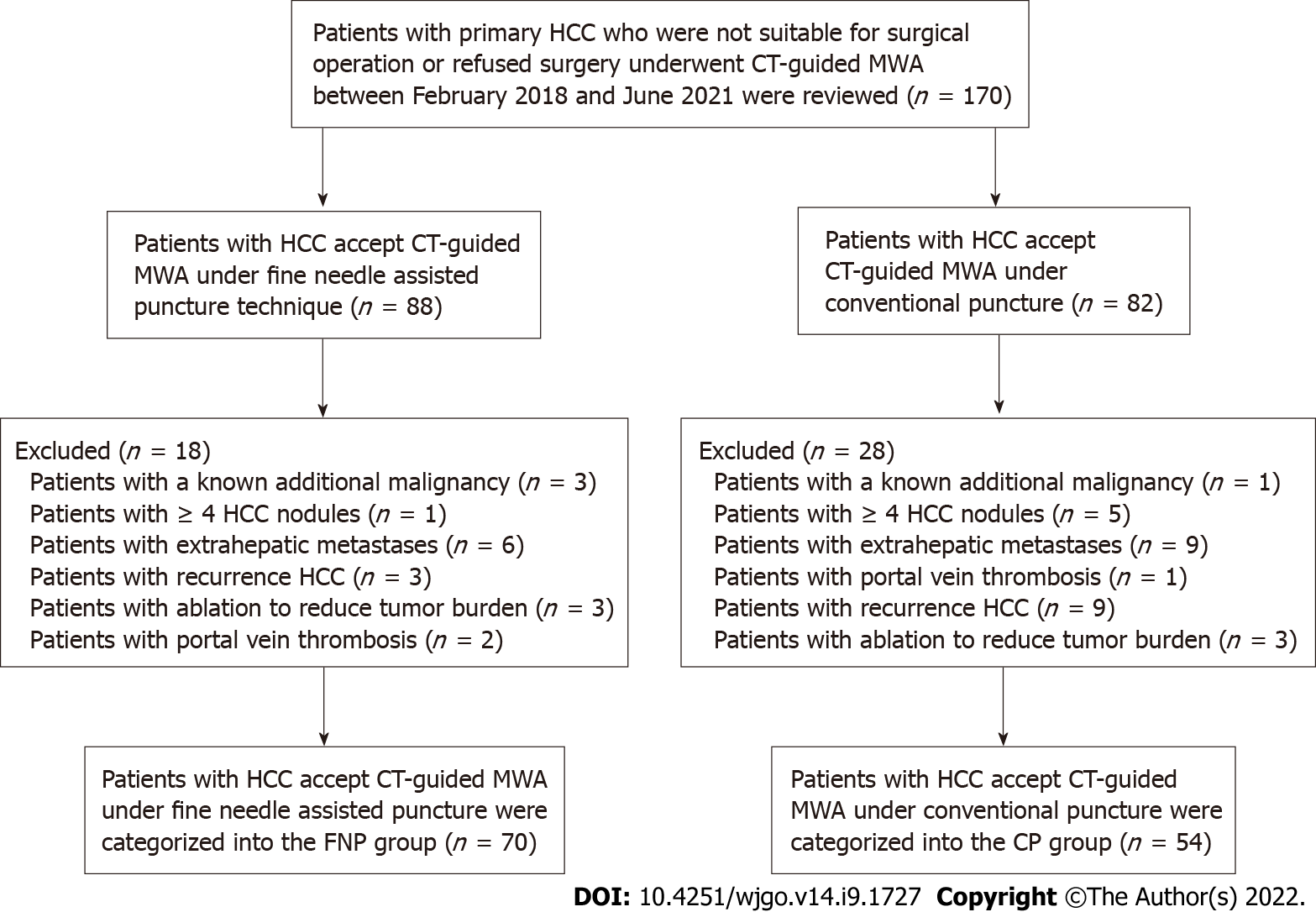
Figure 1 Flow chart showing the selection process of participants for this study.
CT: Computed tomography; CP: Conventional puncture; FNP: Fine needle-assisted puncture positioning; HCC: Hepatocellular carcinoma; MWA: Microwave ablation.
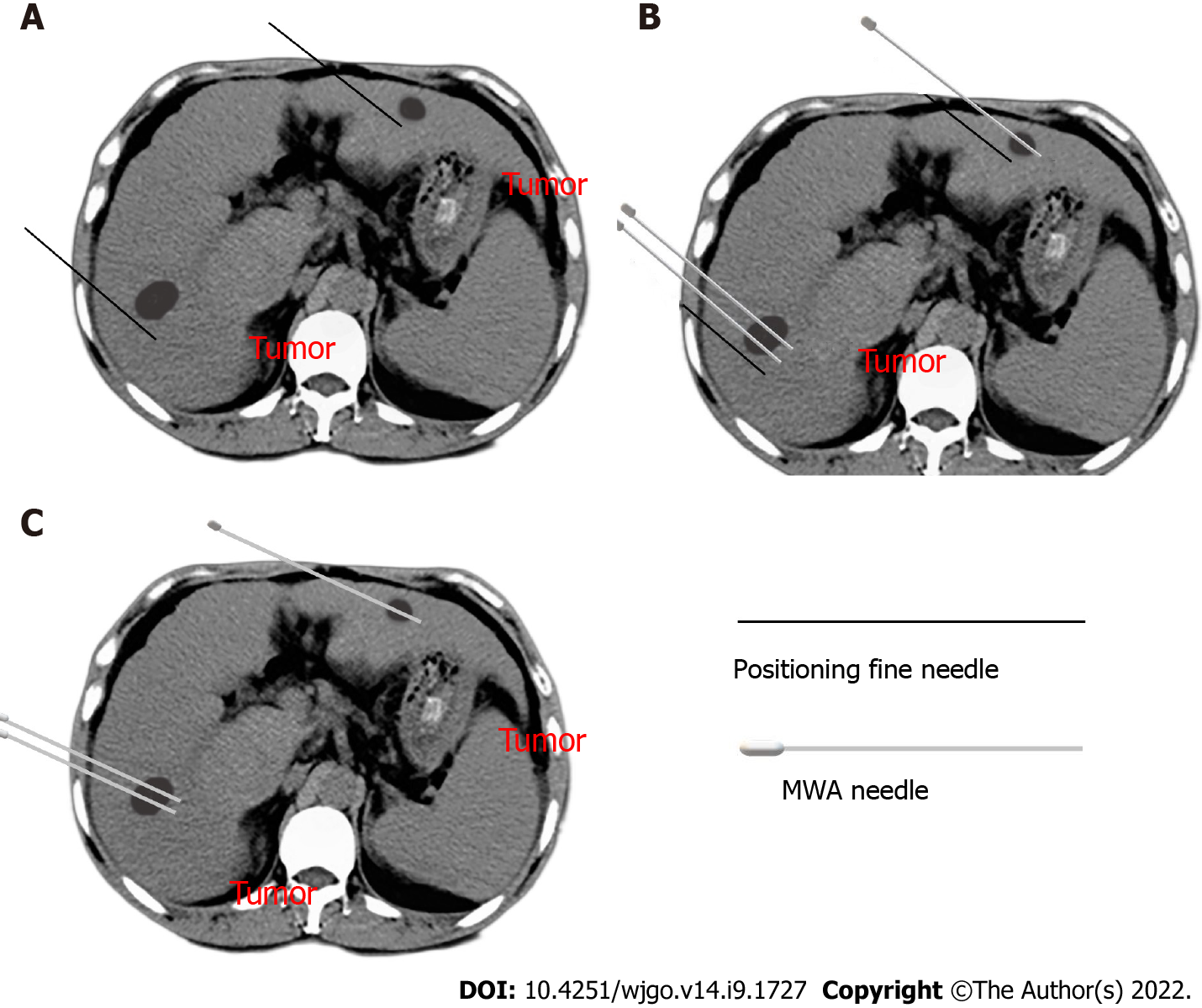
Figure 2 Fine needle-assisted puncture positioning technique in computed tomography-guided microwave ablation.
A: The patient was trained to hold his breath, and then a 21-gauge fine needle was inserted near the tumor nodules; B: A microwave electrode needle was gradually inserted inside the tumor according to the mark of the fine needle while the fine needle served as a breathing indicator; C: The fine needle was pulled out after the electrode needle, consistent with the plan confirmed by computed tomography scanning.
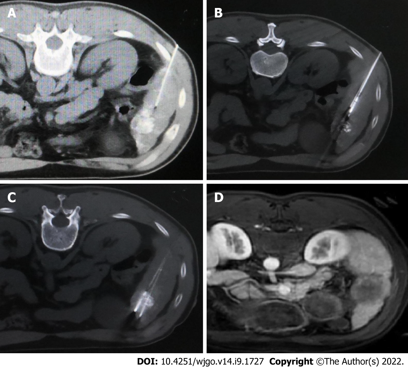
Figure 3 Computed tomography-guided microwave ablation under fine needle-assisted puncture technique for a patient with hepatocellular carcinoma nodule in segment 5 accepted transarterial chemoembolization pre-microwave ablation.
A: A 21-gauge fine needle of 15 cm length was inserted near the tumor nodule after computed tomography scanning before ablation needle puncture; B: A microwave electrode needle was gradually inserted inside the tumor according to the mark of the fine needle; C: The second microwave electrode needle was gradually inserted inside the tumor needle and approximately 1 cm beyond the tumor margin according to the mark of the fine needle; D: The complete ablation was confirmed by magnetic resonance imaging 2 mo post-microwave ablation.
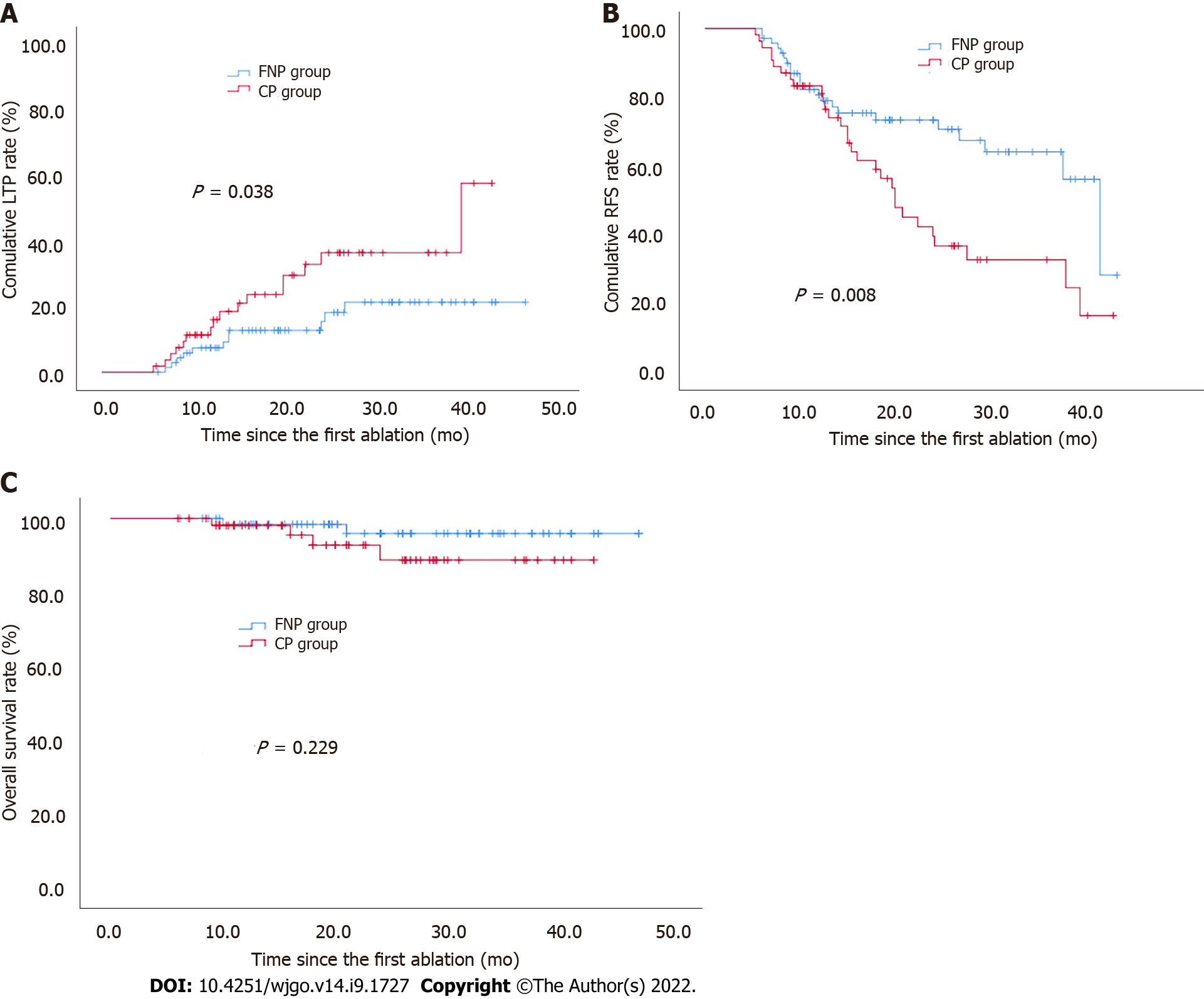
Figure 4 Comparison of cumulative incidence of local tumor progression, recurrence-free survival, and overall survival following post-computed tomography-guided microwave ablation between fine needle-assisted puncture positioning and conventional puncture groups.
A: Local tumor progression (P = 0.038 based on log-rank statistics); B: Recurrence-free survival (P = 0.008 based on log-rank statistics); C: Overall survival (P = 0.229 based on log-rank statistics). CP: Conventional puncture; FNP: Fine needle-assisted puncture positioning; LTP: Local tumor progression; OS: Overall survival; RFS: Recurrence-free survival.
- Citation: Hao MZ, Hu YB, Chen QZ, Chen ZX, Lin HL. Efficacy and safety of computed tomography-guided microwave ablation with fine needle-assisted puncture positioning technique for hepatocellular carcinoma. World J Gastrointest Oncol 2022; 14(9): 1727-1738
- URL: https://www.wjgnet.com/1948-5204/full/v14/i9/1727.htm
- DOI: https://dx.doi.org/10.4251/wjgo.v14.i9.1727


