Copyright
©The Author(s) 2022.
World J Gastrointest Oncol. Aug 15, 2022; 14(8): 1499-1509
Published online Aug 15, 2022. doi: 10.4251/wjgo.v14.i8.1499
Published online Aug 15, 2022. doi: 10.4251/wjgo.v14.i8.1499
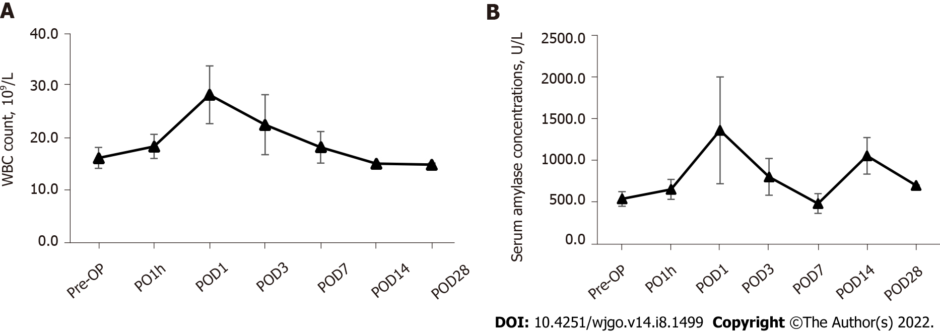
Figure 1 Perioperative changes of white blood cell count and serum amylase.
Both of them were elevated immediately postoperatively (1 h) and peaked after 1 d, then gradually resolved by 4 wk post-ablation. A: White blood cells count; B: Serum amylase. WBC: White blood cell; Pre-OP: Pre-operation; PO1h: Post-operative 1 h; POD: Post-operative day.
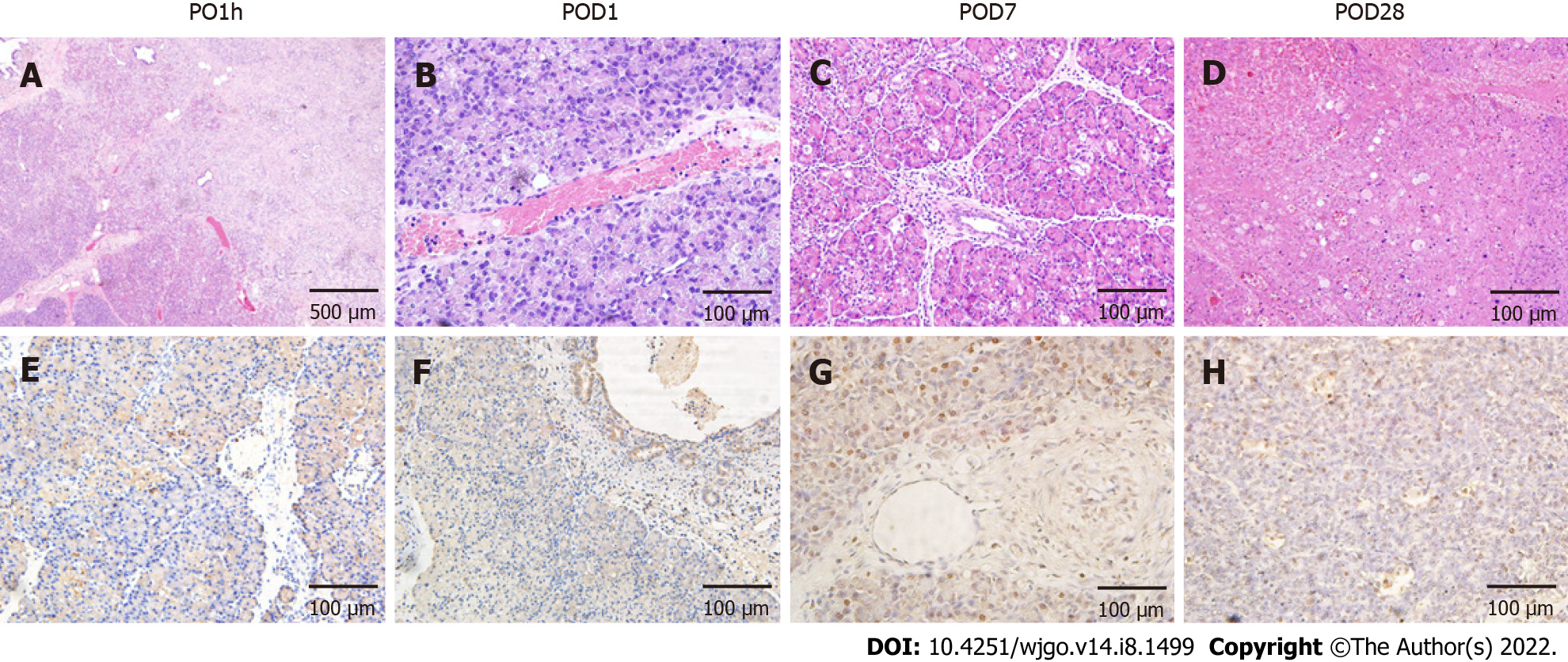
Figure 2 Effects of irreversible electroporation on pancreatic head tissue.
A: Hemotoxylin and eosin staining demonstrated extensive tissue damage in the irreversible electroporation (IRE) ablation zones with clear boundaries between ablation area and nonablation area; B-D: Tissue necrosis and immune cell infiltration were noted up to 4 wk post-IRE with gradual resolution and subsequent mild fibrosis; E-H: Terminal deoxynucleotidyl transferase dUTP nick end labeling (TUNEL) staining revealed that the area centered on the probes in the ablation zone was strongly positive, and apoptotic expression was also seen in pancreatic ductal cells (F) and vascular endothelial cells (G). Scale bar in A = 500 μm. Scale bar in (B-H) = 100 μm. PO1h: Post-operative 1 h; POD: Post-operative day.
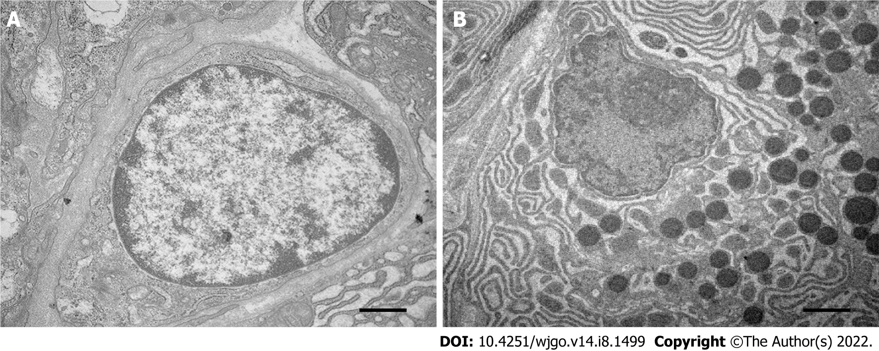
Figure 3 Effects of irreversible electroporation on the ultrastructure of pancreatic acinar cells.
A: Pancreatic acinar cells in the ablation zone showed highly agglutinated and marginalized chromatin, fragmented nucleoli, condensed cytoplasm, and disappearance of rough endoplasmic reticulum; B: Normal control. Scale bar = 500 nm.
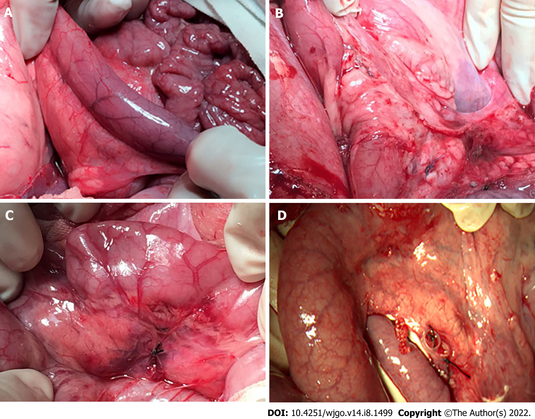
Figure 4 Gross pathology of the duodenum wall after irreversible electroporation.
A: The duodenal wall deepened in color with local congestion and edema 1 h after irreversible electroporation (IRE); B: Postoperative adhesion was observed 24 h after IRE without gastroduodenal obstruction; C and D: The duodenum went back to normal at 1 and 4 wk post-IRE.
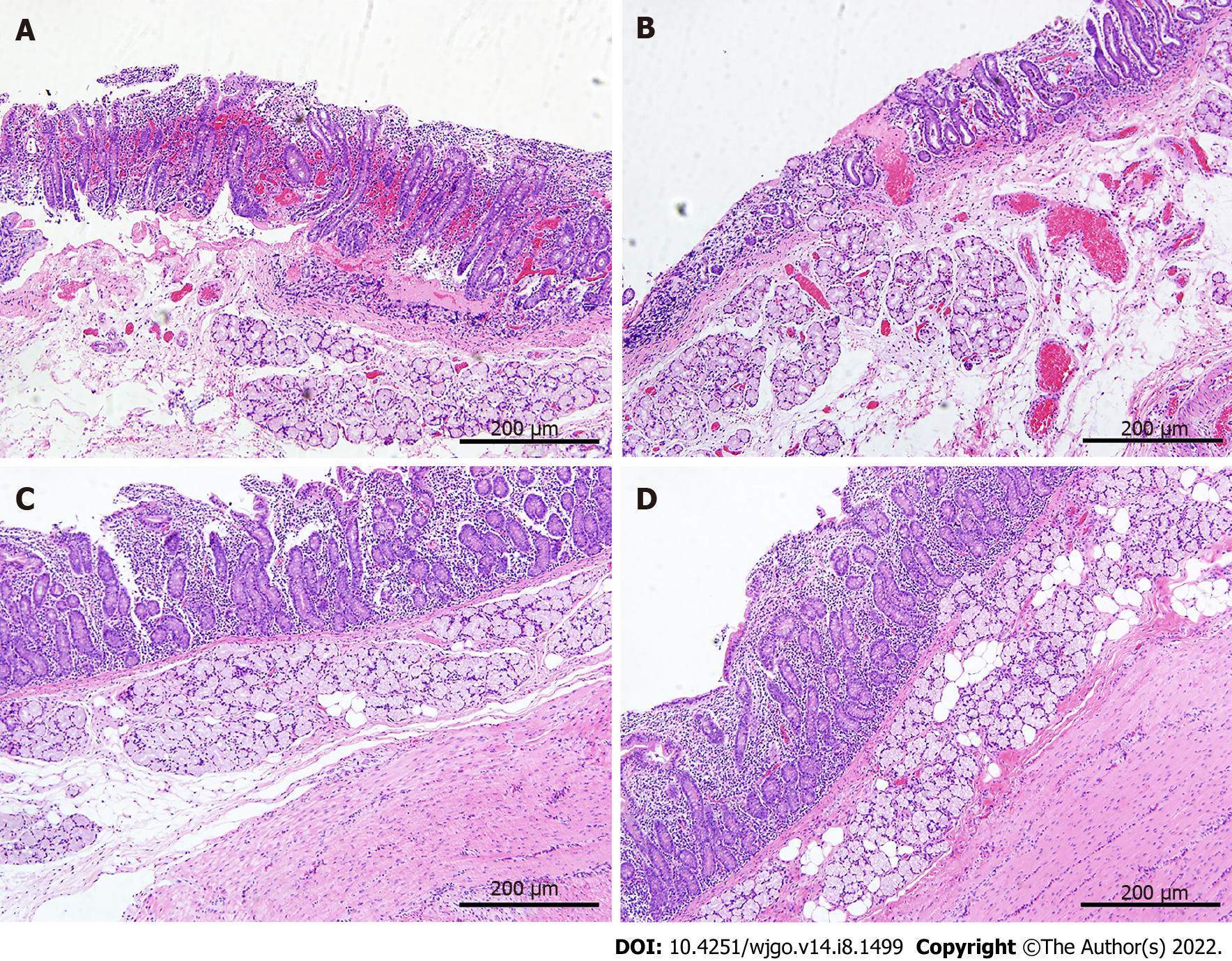
Figure 5 Histopathology of the duodenum wall after irreversible electroporation.
A: The duodenum wall showed necrosis and focal hemorrhage in the mucosa, submucosa, and muscle layers 1 h after irreversible electroporation (IRE); B: The mucosal layer was congested with massive infiltration of inflammatory cells 24 h after IRE; C: On 7 d after IRE new villous structures were observed in the mucosal layer, and immature muscle cells were seen in the muscle layer; D: Twenty-eight days after IRE, the structure of the duodenum appeared intact, and the mucosa layer returned to normal thickness. Scale bars in A-D = 200 μm.
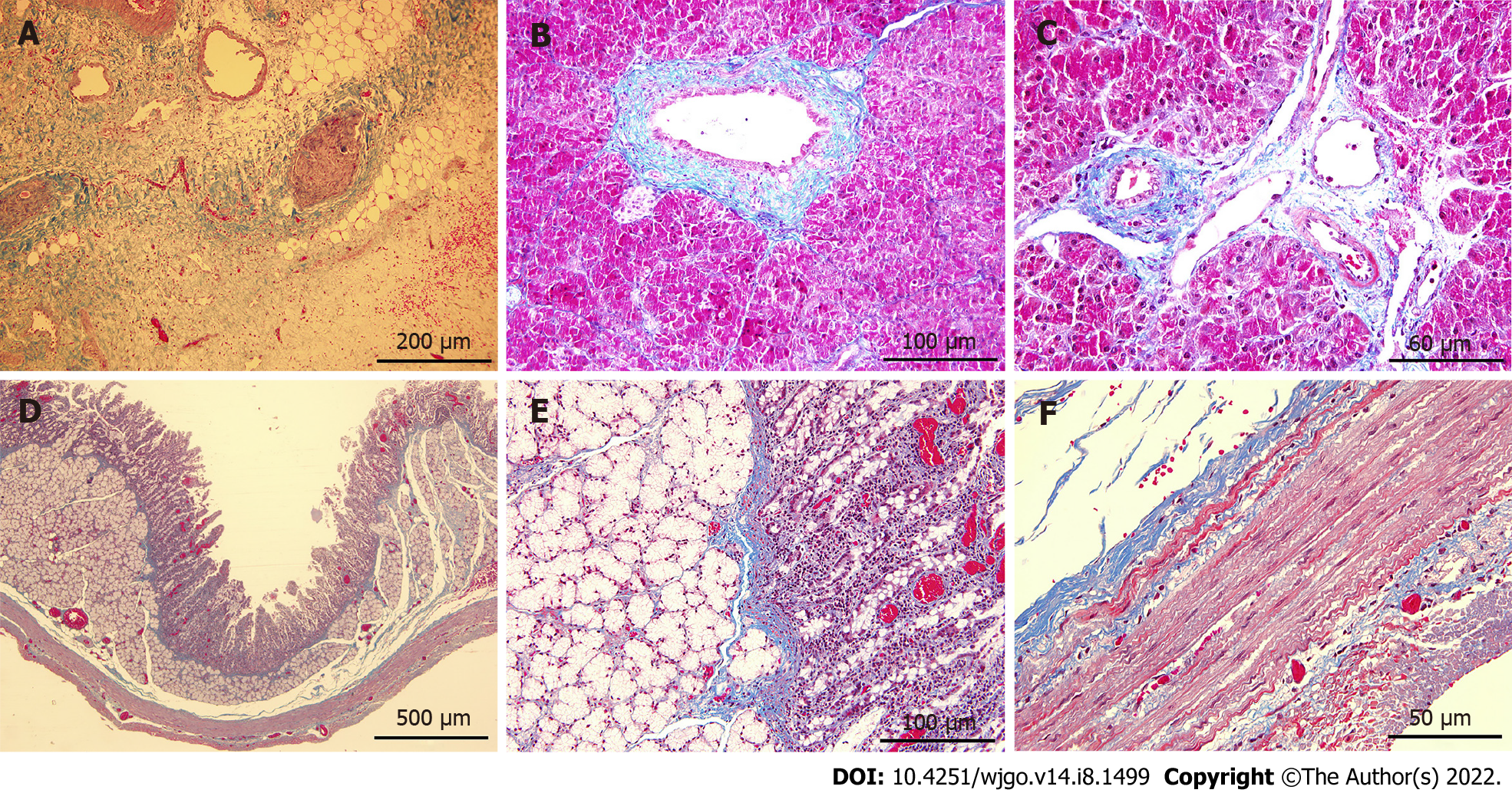
Figure 6 Masson trichrome staining of tissues in the ablation zone.
A-C: Mild fibrosis (blue stained) was observed in pancreatic parenchyma on 7 d post-ablation (A), and the structures of pancreatic ducts (B) and vessels (C) remained intact; D-F: Staining of the duodenum wall showed that the structure of all layers was preserved although minimal injury to the mucosa layer was noted. Scale bars in (A) = 200 μm. Scale bars in (D) = 500 μm. Scale bars in (B and E) = 100 μm. Scale bars in (C and F) = 50 μm.
- Citation: Yan L, Liang B, Feng J, Zhang HY, Chang HS, Liu B, Chen YL. Safety and feasibility of irreversible electroporation for the pancreatic head in a porcine model. World J Gastrointest Oncol 2022; 14(8): 1499-1509
- URL: https://www.wjgnet.com/1948-5204/full/v14/i8/1499.htm
- DOI: https://dx.doi.org/10.4251/wjgo.v14.i8.1499


