Copyright
©The Author(s) 2022.
World J Gastrointest Oncol. Aug 15, 2022; 14(8): 1429-1445
Published online Aug 15, 2022. doi: 10.4251/wjgo.v14.i8.1429
Published online Aug 15, 2022. doi: 10.4251/wjgo.v14.i8.1429
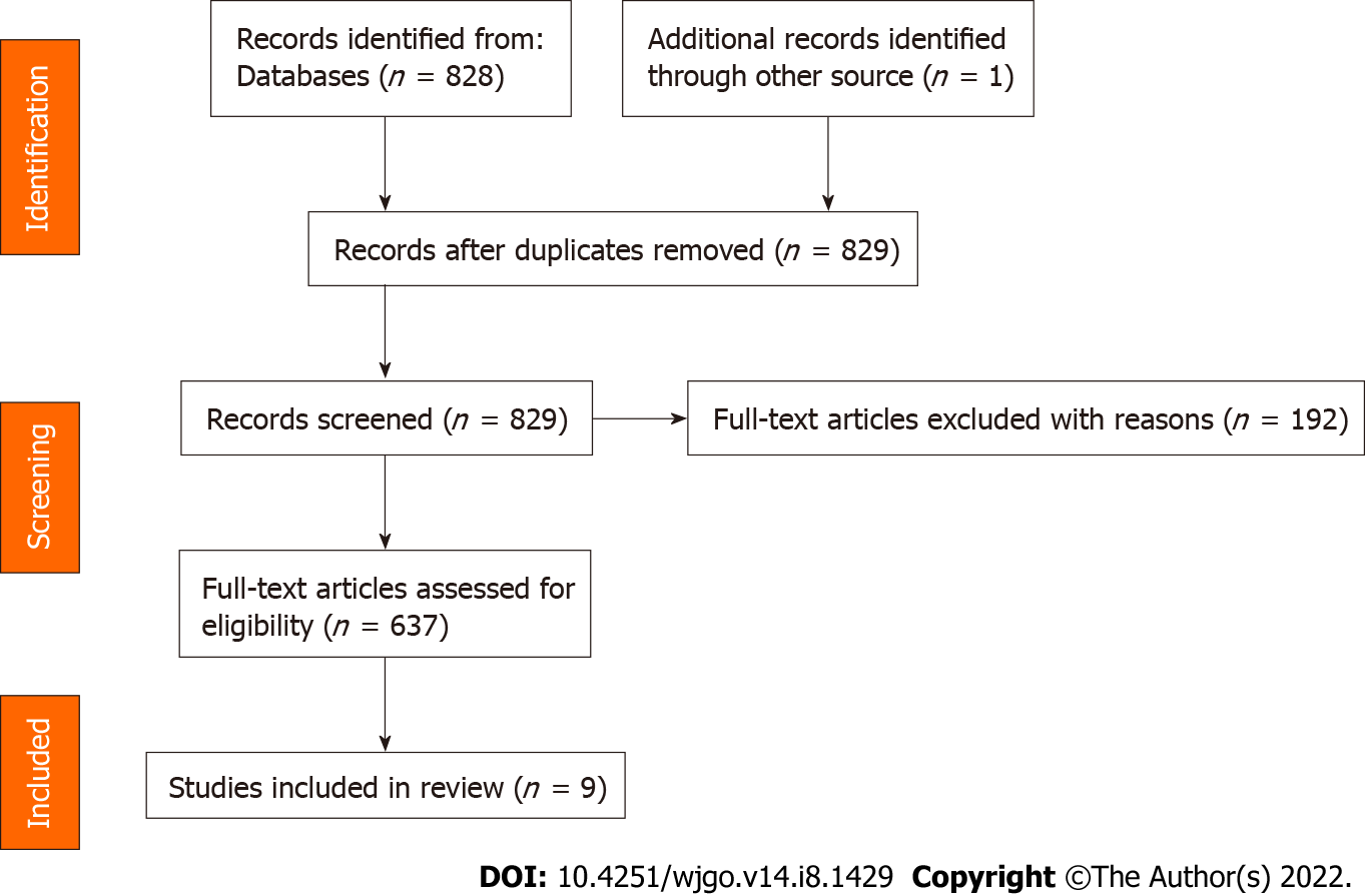
Figure 1 Preferred Reporting Items for Systematic Reviews and Meta-Analyses (2020) flow diagram.
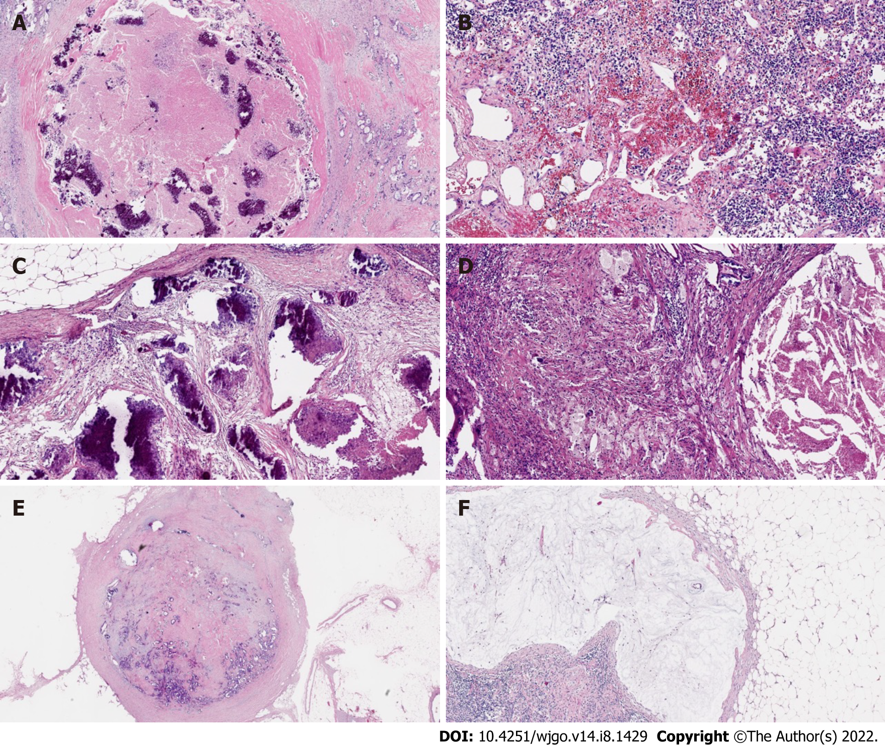
Figure 2 Example of modes of lymph node tumor regression.
A: Necrosis; B: Hemorrhage, nodular; C: Fibrosis; D: Foamy histiocytes; E: Residual cancer cells; F: Pools of mucin.
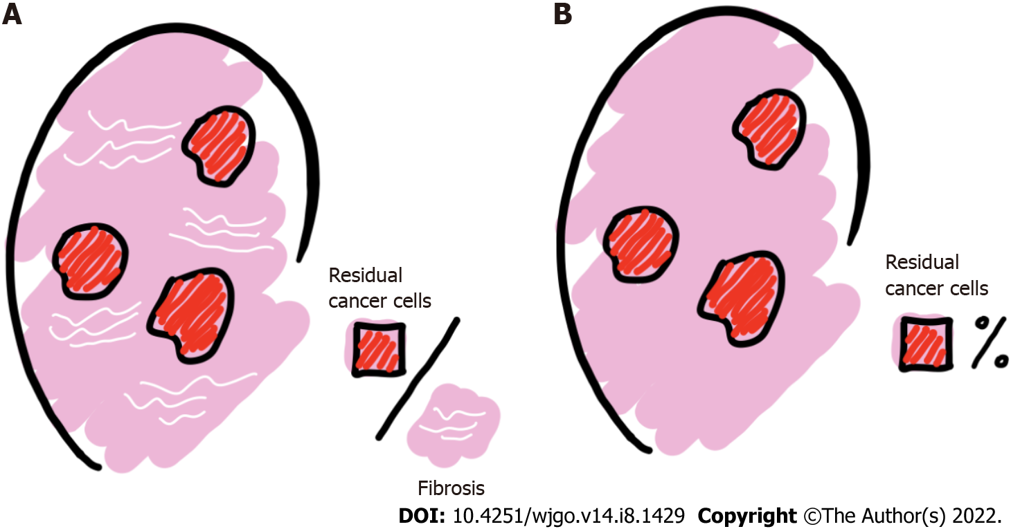
Figure 3 Principles of lymph node regression grade assessment.
A: Ratio of residual cancer cells to fibrosis; B: Percentage of residual cancer cells in the lymph nodes.
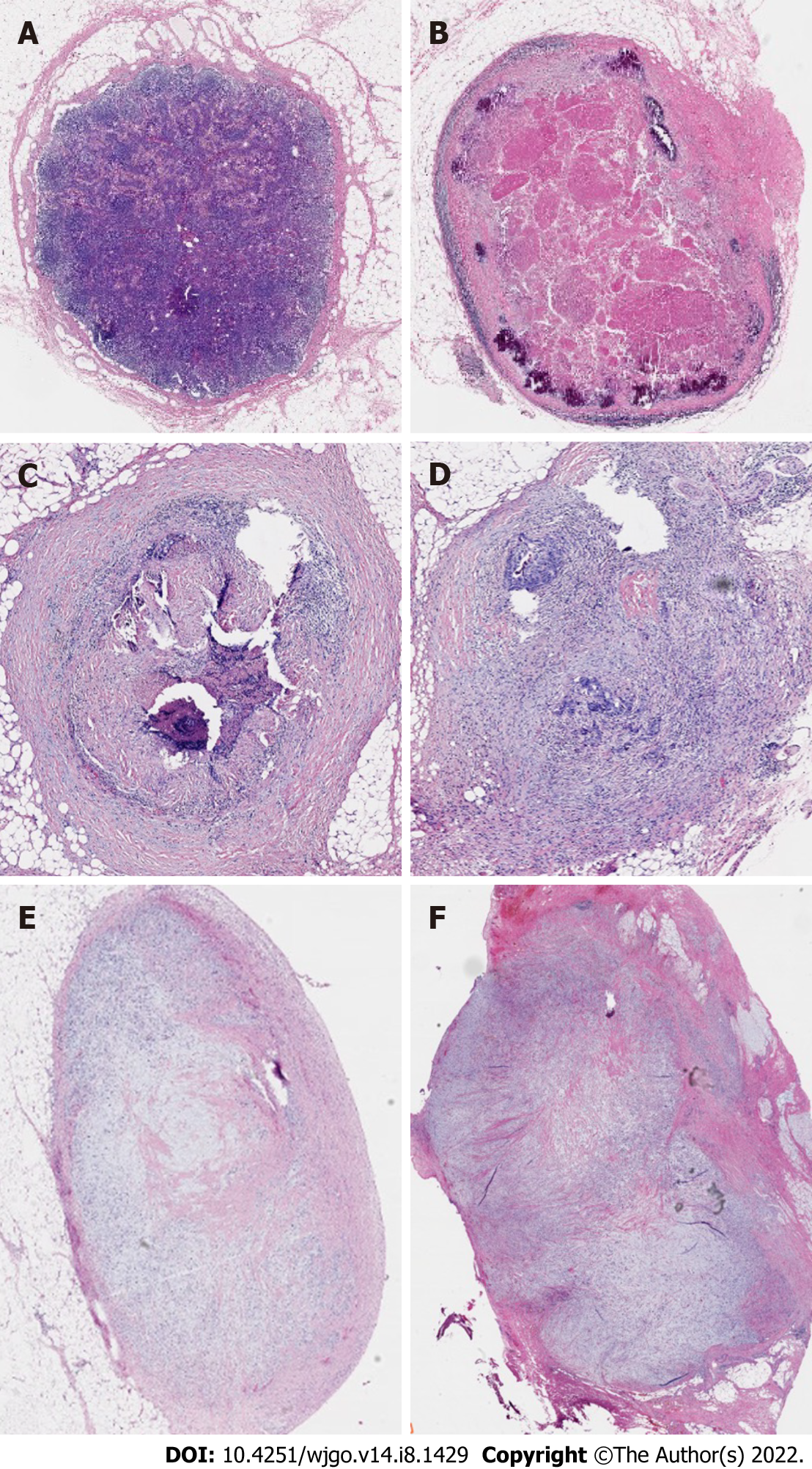
Figure 4 Examples of lymph node regression grades according to Mirbagheri et al[28] (10 ×).
A: Lymph node regression grade (LRG) 0: Normal lymph node; B: LRG1: 100% fibrosis, no residual cancer; C: LRG2: 75%-100% fibrosis, 0-25% cancer; D: LRG3: 50%-75% fibrosis, 25%-50% cancer; E: LRG4: 25%-50% fibrosis, 50%-75% cancer; F: LRG5: 0-25% fibrosis, 75%-100% cancer.
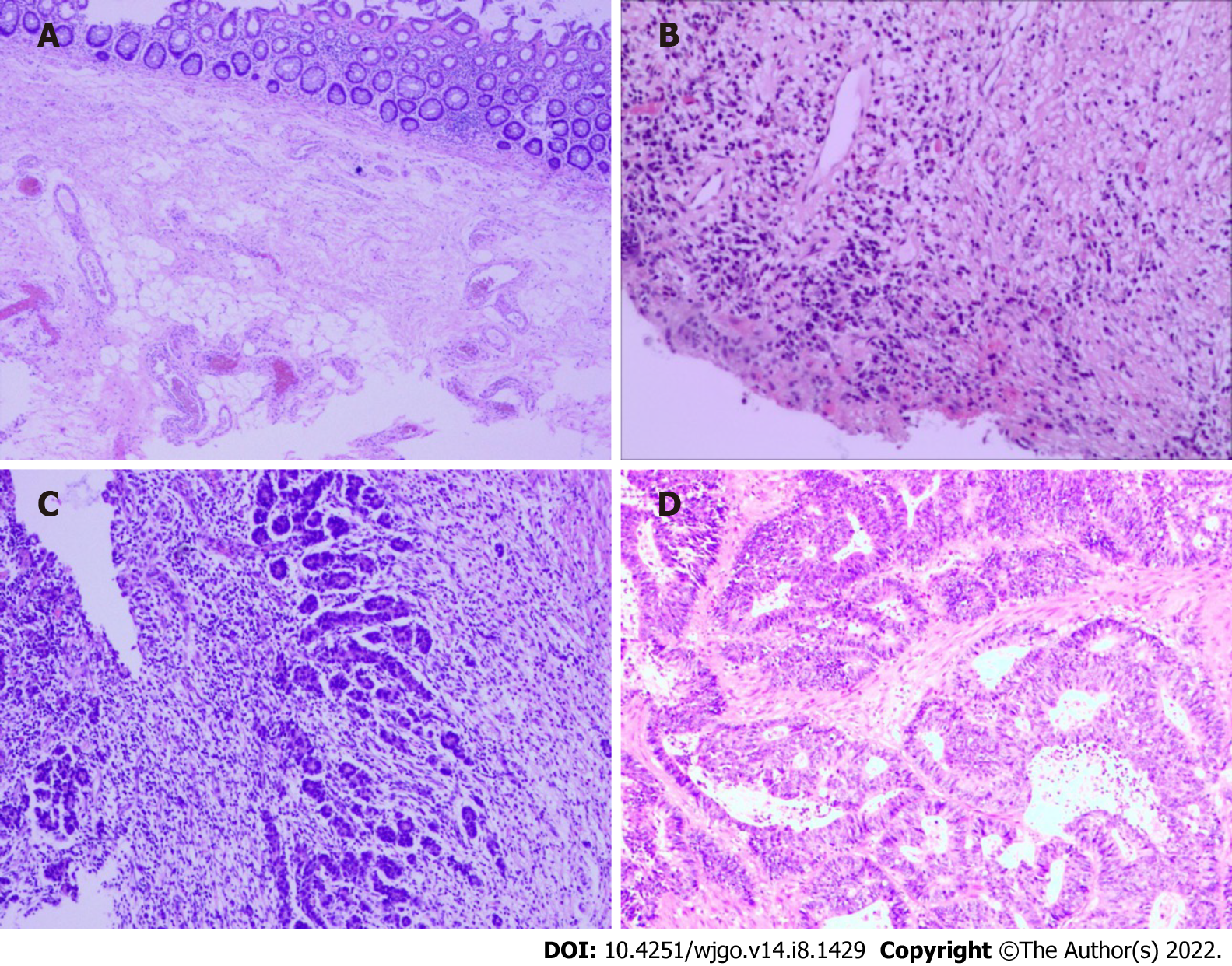
Figure 5 Examples of tumor regression grades according to American Joint Committee on Cancer.
A: Tumor regression grade (TRG) 0: complete-no viable cells present; B: TRG1: moderate-single cells/small groups of cancer cells; C: TRG2: minimal-residual cancer outgrown by fibrosis; D: TRG3: poor-minimal or no tumor cell death, extensive residual cancer.
- Citation: He L, Xiao J, Zheng P, Zhong L, Peng Q. Lymph node regression grading of locally advanced rectal cancer treated with neoadjuvant chemoradiotherapy. World J Gastrointest Oncol 2022; 14(8): 1429-1445
- URL: https://www.wjgnet.com/1948-5204/full/v14/i8/1429.htm
- DOI: https://dx.doi.org/10.4251/wjgo.v14.i8.1429


