Copyright
©The Author(s) 2022.
World J Gastrointest Oncol. May 15, 2022; 14(5): 959-972
Published online May 15, 2022. doi: 10.4251/wjgo.v14.i5.959
Published online May 15, 2022. doi: 10.4251/wjgo.v14.i5.959
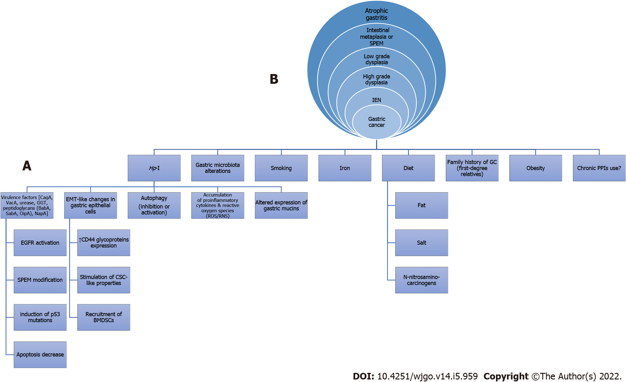
Figure 1 Possible mechanisms involved (A) in the etiology of non-cardiac gastric cancer (intestinal type) resulting in the classical cascade of Correa histopathological precancerous lesions (B) as seen in an upper gastrointestinal endoscopy.
Hp-I: Helicobacter pylori infection; GC: Gastric cancer; PPIs: Proton pump inhibitors; CagA: Cytotoxin-associated gene A; VacA: Vacuolating cytotoxin A; GGT: γ-glutamyl transpeptidase; BabA: Blood-group-antigen-binding adhesin; SabA: Sialic acid-binding adhesin; OipA: Outer inflammatory protein; NapA: Neutrophil activation protein A; EMT: Epithelial-mesenchymal transition; ROS/RNS: Reactive oxygen species/Reactive nitrogen species; EGFR: Epidermal growth factor receptor; SPEM: Spasmolytic polypeptide-expressing metaplasia; CSC: Cancer stem cell; BMDSCs: Bone marrow-derived stem cells; IEN: Intraepithelial neoplasia.
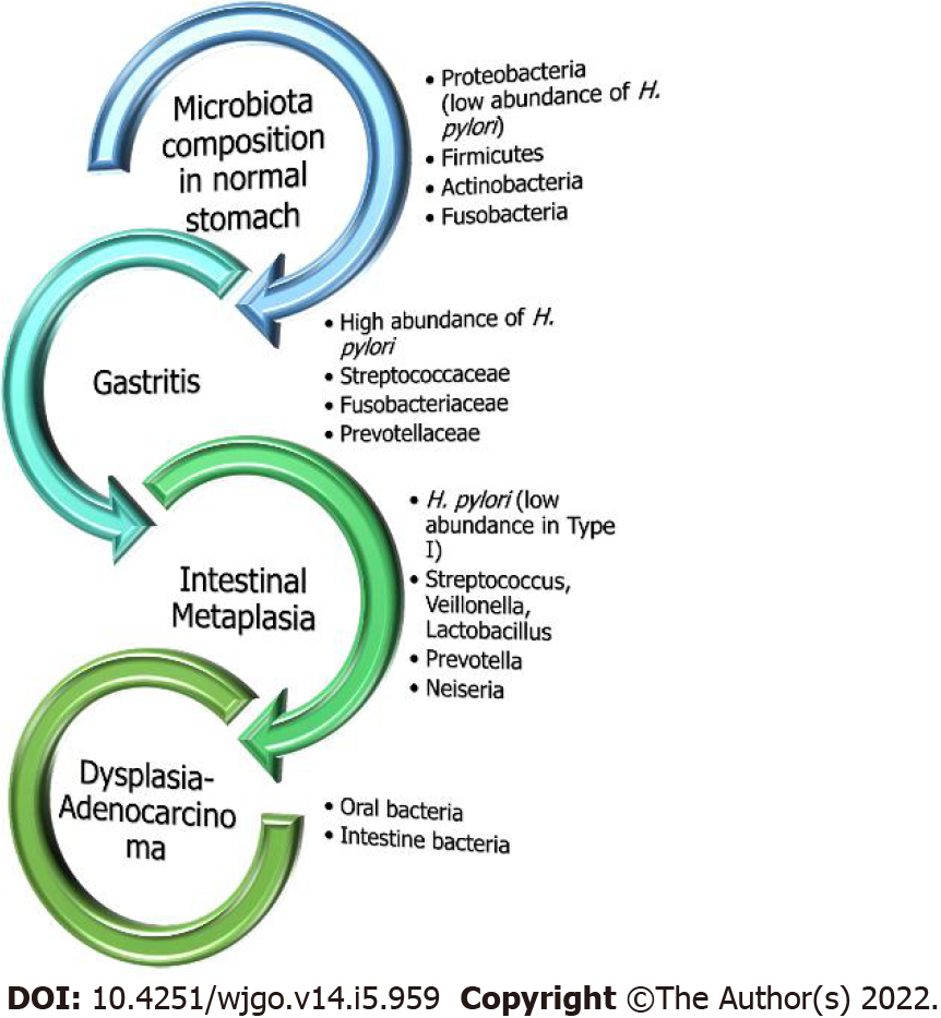
Figure 2 Gastric microbial composition in the healthy and diseased stomach.
Under normal healthy conditions without evidence of excessive inflammation, Helicobacter pylori (H. pylori) exists in very low abundance. On the contrary, in chronic gastritis, H. pylori is the predominant bacteria with the presence of other microorganisms as well but at lower rates. However, as the sequalae of carcinogenesis moves towards malignancy, oral or intestinal-type pathogens exclusively predominate.
- Citation: Liatsos C, Papaefthymiou A, Kyriakos N, Galanopoulos M, Doulberis M, Giakoumis M, Petridou E, Mavrogiannis C, Rokkas T, Kountouras J. Helicobacter pylori, gastric microbiota and gastric cancer relationship: Unrolling the tangle. World J Gastrointest Oncol 2022; 14(5): 959-972
- URL: https://www.wjgnet.com/1948-5204/full/v14/i5/959.htm
- DOI: https://dx.doi.org/10.4251/wjgo.v14.i5.959


