Copyright
©The Author(s) 2022.
World J Gastrointest Oncol. Nov 15, 2022; 14(11): 2295-2301
Published online Nov 15, 2022. doi: 10.4251/wjgo.v14.i11.2295
Published online Nov 15, 2022. doi: 10.4251/wjgo.v14.i11.2295
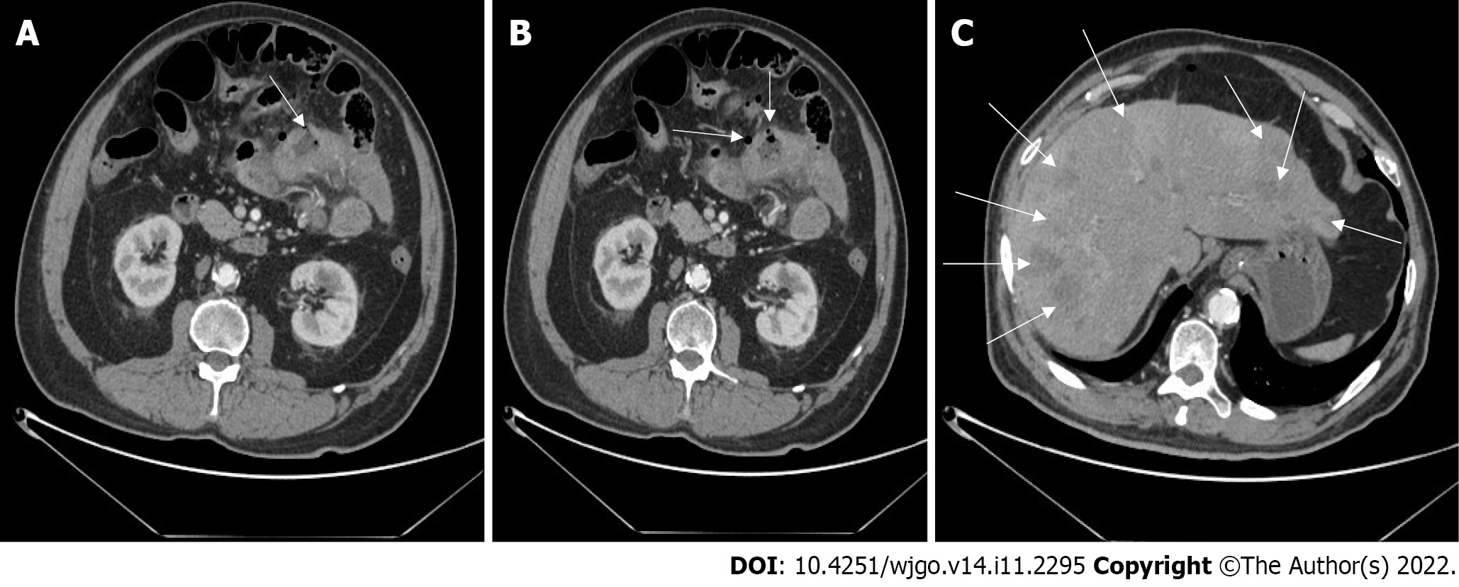
Figure 1 Abdominal computed tomography images.
A: Uneven thickening of the small intestinal wall with viscus perforation (arrow); B: Free gas outside the intestine but in the abdomen (arrows); C: The liver parenchyma had multiple round low-density nodules of varying sizes (arrows).
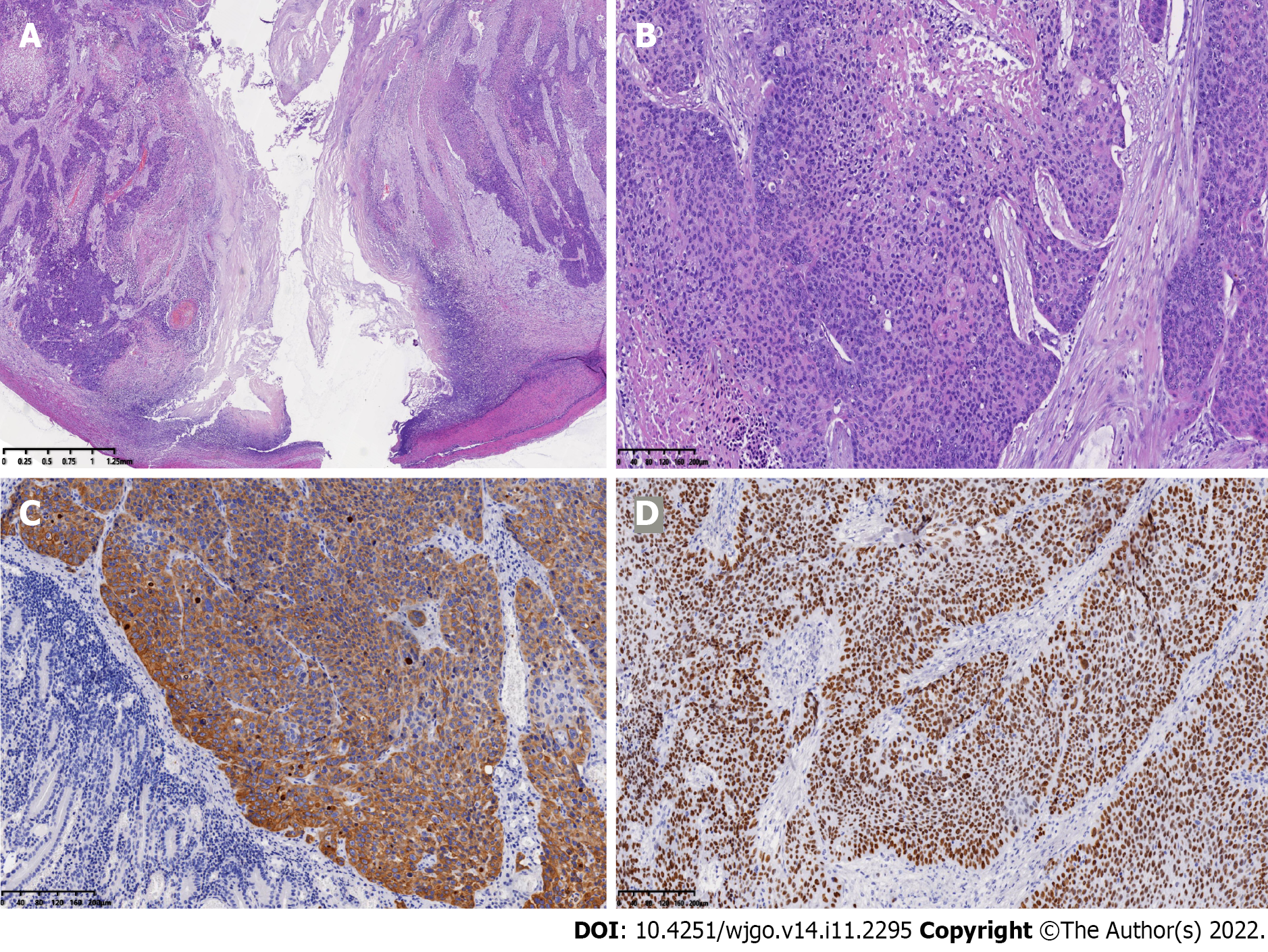
Figure 2 Pathological and immunohistochemical findings.
A and B: Pathological findings from surgical specimen. The lesion showed a serosal penetration (A), with diffuse and nested growth of tumor cells and intracellular dyskeratosis being visible (B); C and D: Immunochemistry demonstrated that the staining for cytokeratin-5/6 and antioncogene P40 was strongly positive.
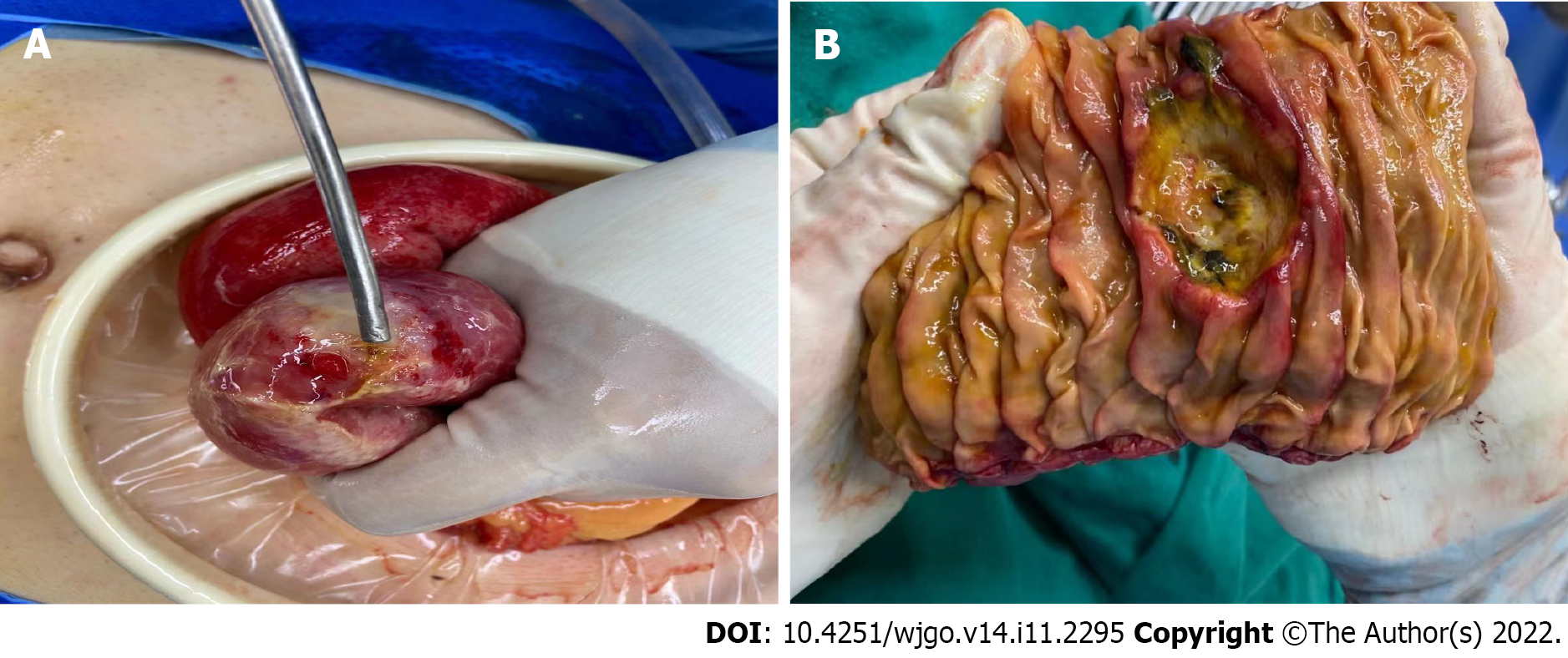
Figure 3 Exploratory laparotomy results.
A: The perforation of a jejunal segment with a purulent surface was noted; B: The enteric cavity showed a deep ulcer lesion.
- Citation: Xiao L, Sun L, Zhang JX, Pan YS. Rare squamous cell carcinoma of the jejunum causing perforated peritonitis: A case report. World J Gastrointest Oncol 2022; 14(11): 2295-2301
- URL: https://www.wjgnet.com/1948-5204/full/v14/i11/2295.htm
- DOI: https://dx.doi.org/10.4251/wjgo.v14.i11.2295


