Copyright
©The Author(s) 2022.
World J Gastrointest Oncol. Oct 15, 2022; 14(10): 1949-1967
Published online Oct 15, 2022. doi: 10.4251/wjgo.v14.i10.1949
Published online Oct 15, 2022. doi: 10.4251/wjgo.v14.i10.1949
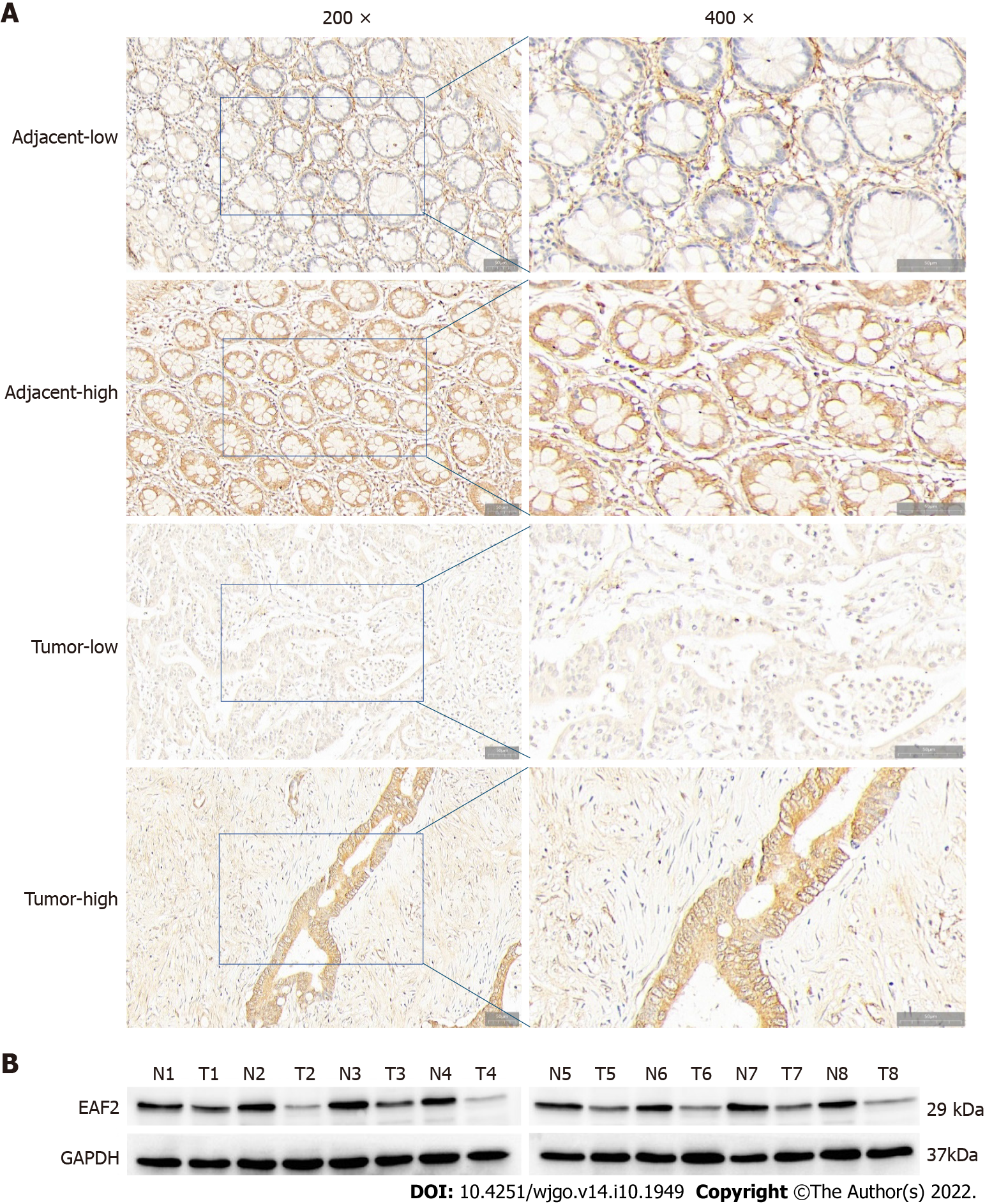
Figure 1 ELL-associated factor 2 protein is decreased in colorectal cancer tissue.
A: Representative immunohistochemical staining for ELL-associated factor 2 (EAF2) in colorectal adenocarcinoma tissue and adjacent tissue (magnification, 200 × and 400 ×); B: western blot showing lower EAF2 level in colorectal adenocarcinoma fresh tissue than in corresponding para-cancerous tissue (T, colorectal adenocarcinoma tissue; N, adjacent non-tumor tissue). EAF2: ELL-associated factor 2; GAPDH: Glyceraldehyde-3-phosphate dehydrogenase.
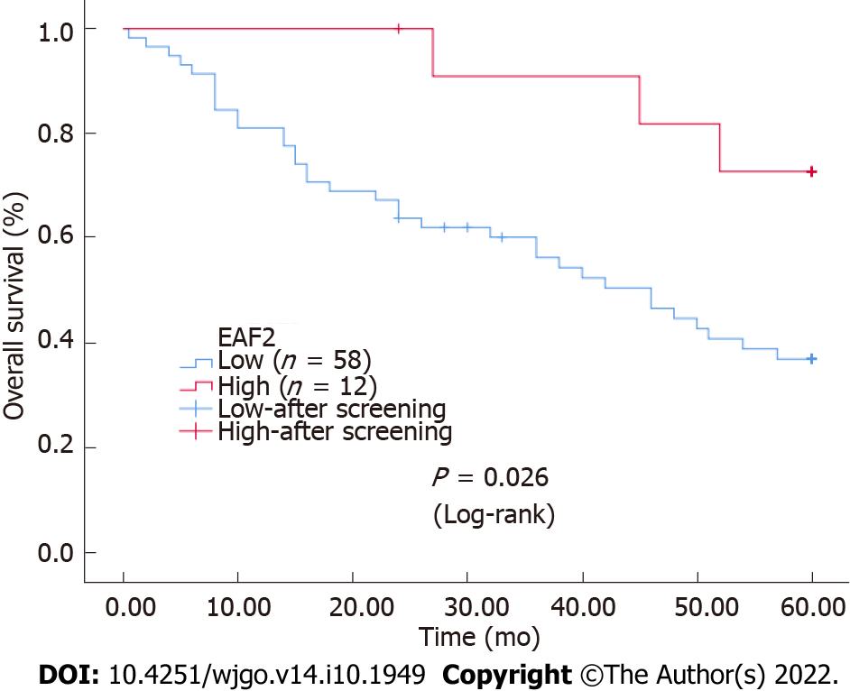
Figure 2 Kaplan-Meier survival curves for ELL-associated factor 2 expression in colorectal cancer tissue.
The survival rate of the group with high ELL-associated factor 2 (EAF2) levels was higher than that of the group with low EAF2 levels. EAF2: ELL-associated factor 2.
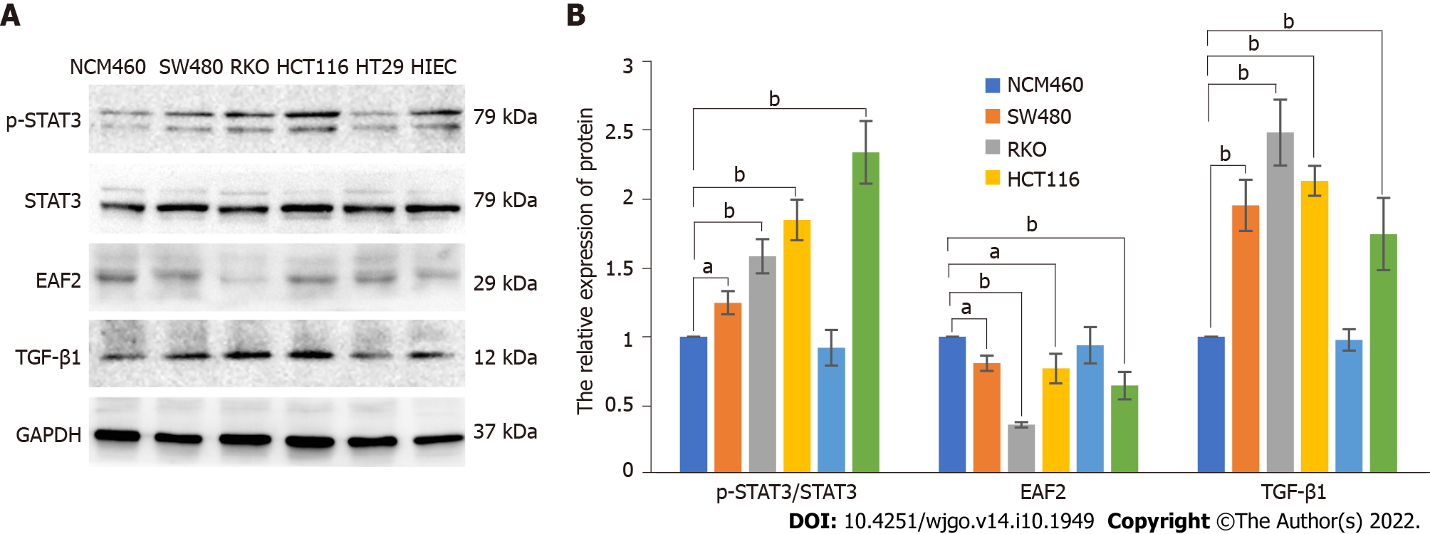
Figure 3 Protein levels of ELL-associated factor 2, phosphorylated-signal transducer and activator of transcription 3, and transforming growth factor-β1 in colorectal cancer cell lines (SW480, RKO, HCT116, HT29, and HIEC) and normal colorectal epithelial cells (NCM460) by western blot assay.
A: Western blot results of proteins in in colorectal cancer cell lines (SW480, RKO, HCT116, HT29, and HIEC) and normal colorectal epithelial cells (NCM460); B: Relative expression of the proteins. The data are shown as the mean ± SD. aP < 0.05, bP < 0.001. p-STAT3: Phosphorylated-signal transducer and activator of transcription 3; TGF-β1: Transforming growth factor-β1; EAF2: ELL-associated factor 2; GAPDH: Glyceraldehyde-3-phosphate dehydrogenase.
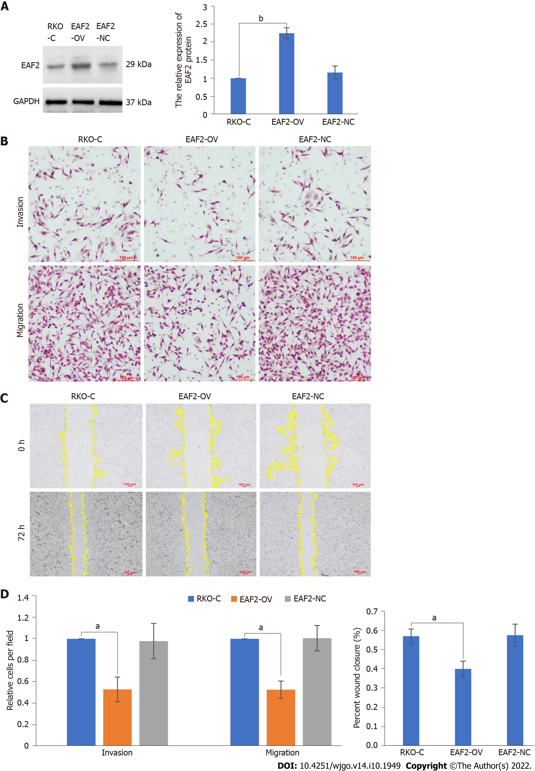
Figure 4 Overexpression of ELL-associated factor 2 inhibits the invasion and migration of RKO cells.
A: Protein levels of ELL-associated factor 2 (EAF2) in RKO cells transfected with EAF2 overexpression vector by western blot assay; B: Invasion and migration abilities evaluated by transwell assay; C: Migration ability analyzed by wound healing assay after 72 h; D: Migration and invasion abilities of RKO cells measured using ImageJ software. The data are shown as the mean ± SD. aP < 0.001; bP < 0.05. RKO-C: The group of RKO control cells; EAF2-OV: The group of RKO cells transfected with ELL-associated factor 2 overexpression vector; EAF2-NC: The group of RKO cells transfected empty vector; EAF2: ELL-associated factor 2; GAPDH: Glyceraldehyde-3-phosphate dehydrogenase.
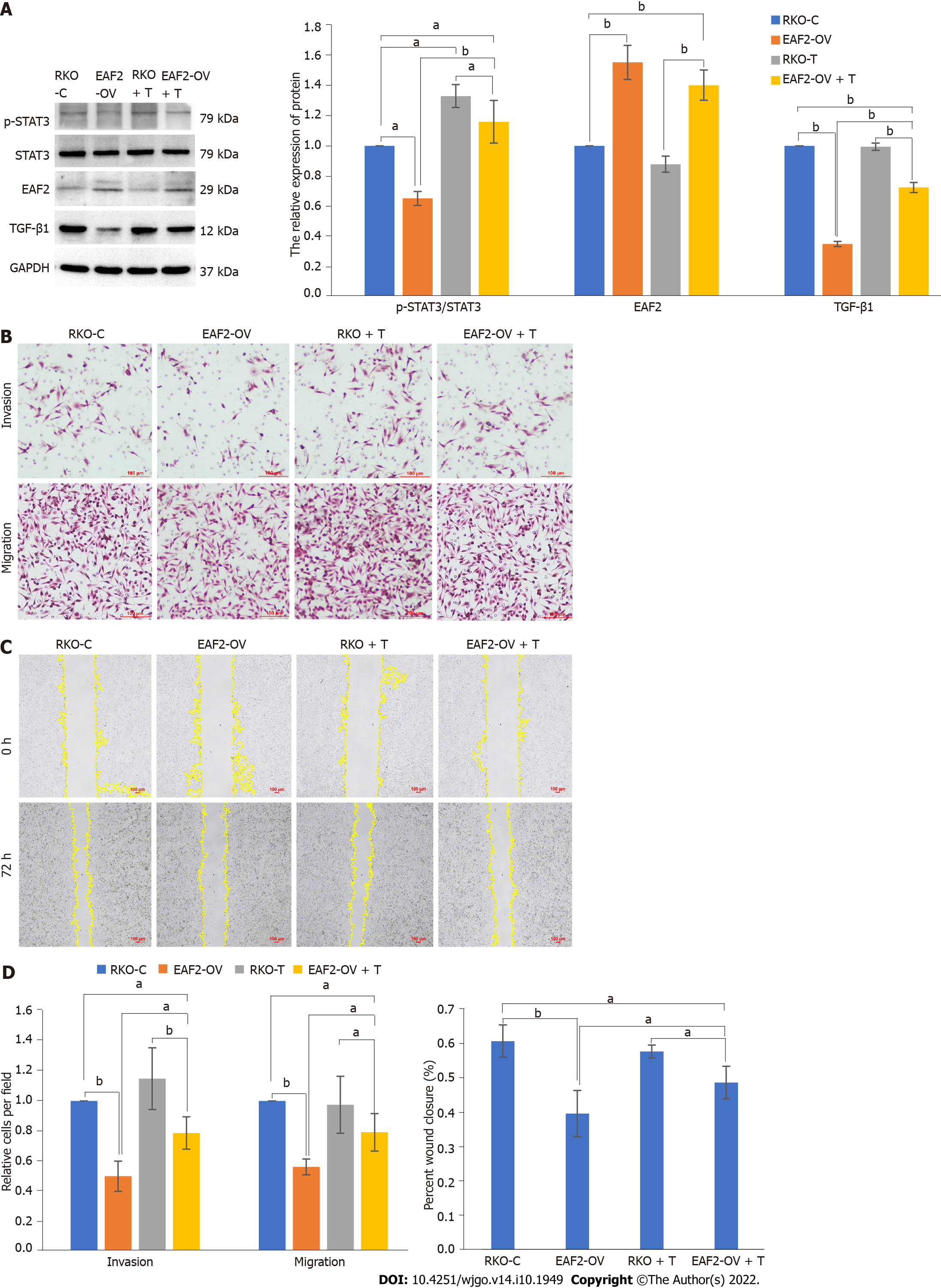
Figure 5 Overexpression of ELL-associated factor 2 suppresses the activity of the signal transducer and activator of transcription 3/transforming growth factor-β1 pathway to inhibit the invasion and migration of RKO cells.
A: Overexpression of ELL-associated factor 2 (EAF2) inhibited the signal transducer and activator of transcription 3/transforming growth factor-β1 (STAT3/TGF-β1) pathway measured by western blot assay. And TGF-β1 recombinant protein abolished EAF2 overexpression-reduced the phosphorylation (Tyr705) of STAT3 protein; B: Invasion and migration abilities evaluated by transwelll assay; C: Migration ability analyzed by wound healing assay after 72 h; D: Migration and invasion abilities of RKO cells measured using ImageJ software. The data are shown as the mean ± SD. aP < 0.05, bP < 0.001. RKO-C: The group of RKO control cells; EAF2-OV: The group of RKO cells transfected with ELL-associated factor 2 overexpression vector; RKO + T: The group of RKO cells treated with 5 ng/mL transforming growth factor-β1 recombinant protein for 24 h; EAF2-OV + T: The group of RKO cells were transfected with EAF2-OV plasmid for 24 h and then cultured with 5 ng/mL transforming growth factor-β1 recombinant protein for 24 h; p-STAT3: Phosphorylated-signal transducer and activator of transcription 3; TGF-β1: Transforming growth factor-β1; EAF2: ELL-associated factor 2; GAPDH: Glyceraldehyde-3-phosphate dehydrogenase.
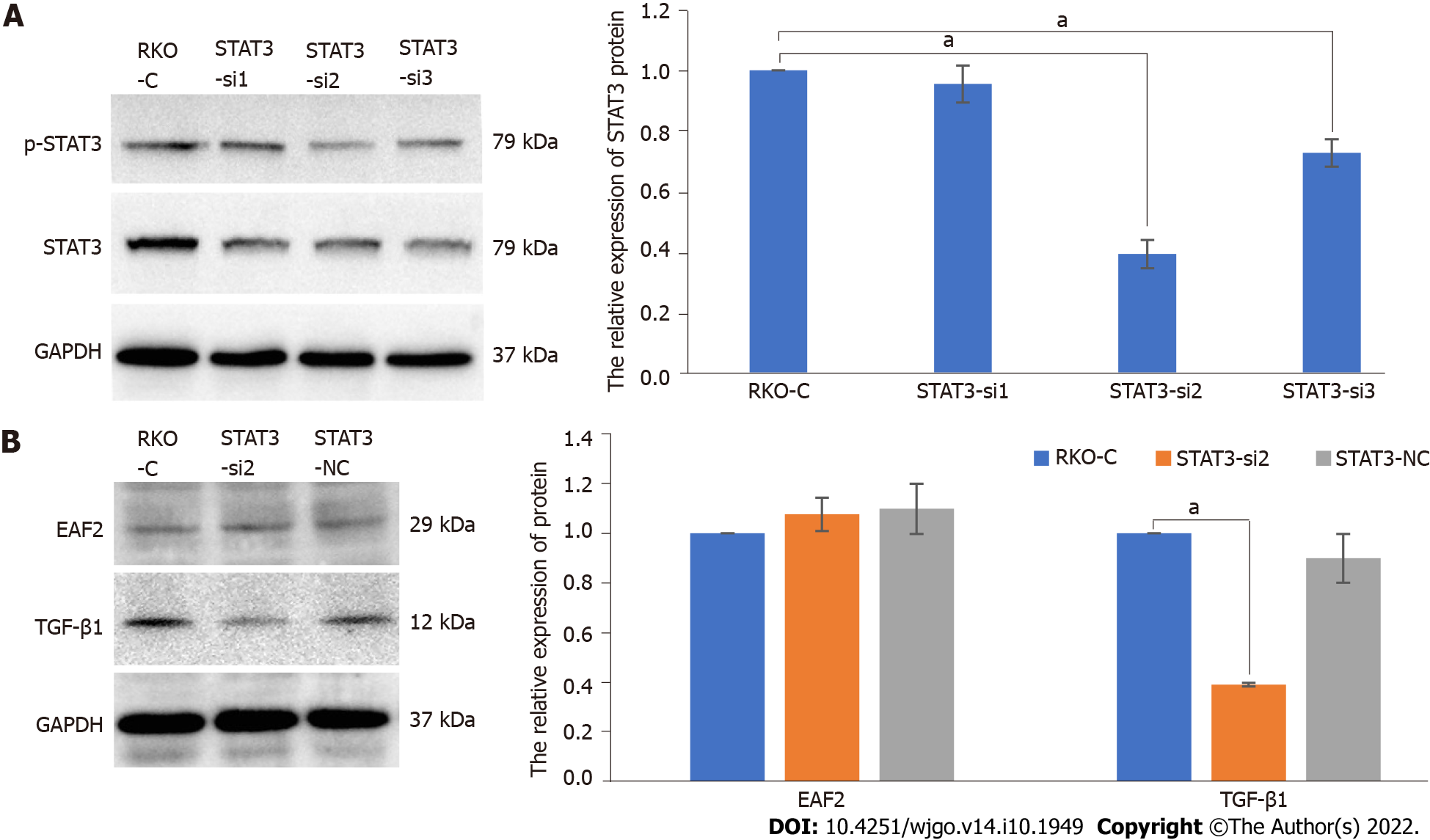
Figure 6 Silencing signal transducer and activator of transcription 3 decreases transforming growth factor-β1 protein expression in RKO cells.
A: Protein levels of phosphorylated-signal transducer and activator of transcription 3 (p-STAT3) in RKO cells transfected with STAT3 siRNA plasmid by western blot assay; B: Knockdown of STAT3 decreased the levels of transforming growth factor-β1 protein but not ELL-associated factor 2 protein in RKO cells. The data are shown as the mean ± SD. aP < 0.01. RKO-C: The group of RKO control cells; STAT3-si: The group of RKO cells transfected with signal transducer and activator of transcription 3 siRNA plasmid. STAT3-NC: The group of RKO cells transfected empty vector; p-STAT3: Phosphorylated-signal transducer and activator of transcription 3; TGF-β1: Transforming growth factor-β1; EAF2: ELL-associated factor 2; GAPDH: Glyceraldehyde-3-phosphate dehydrogenase.
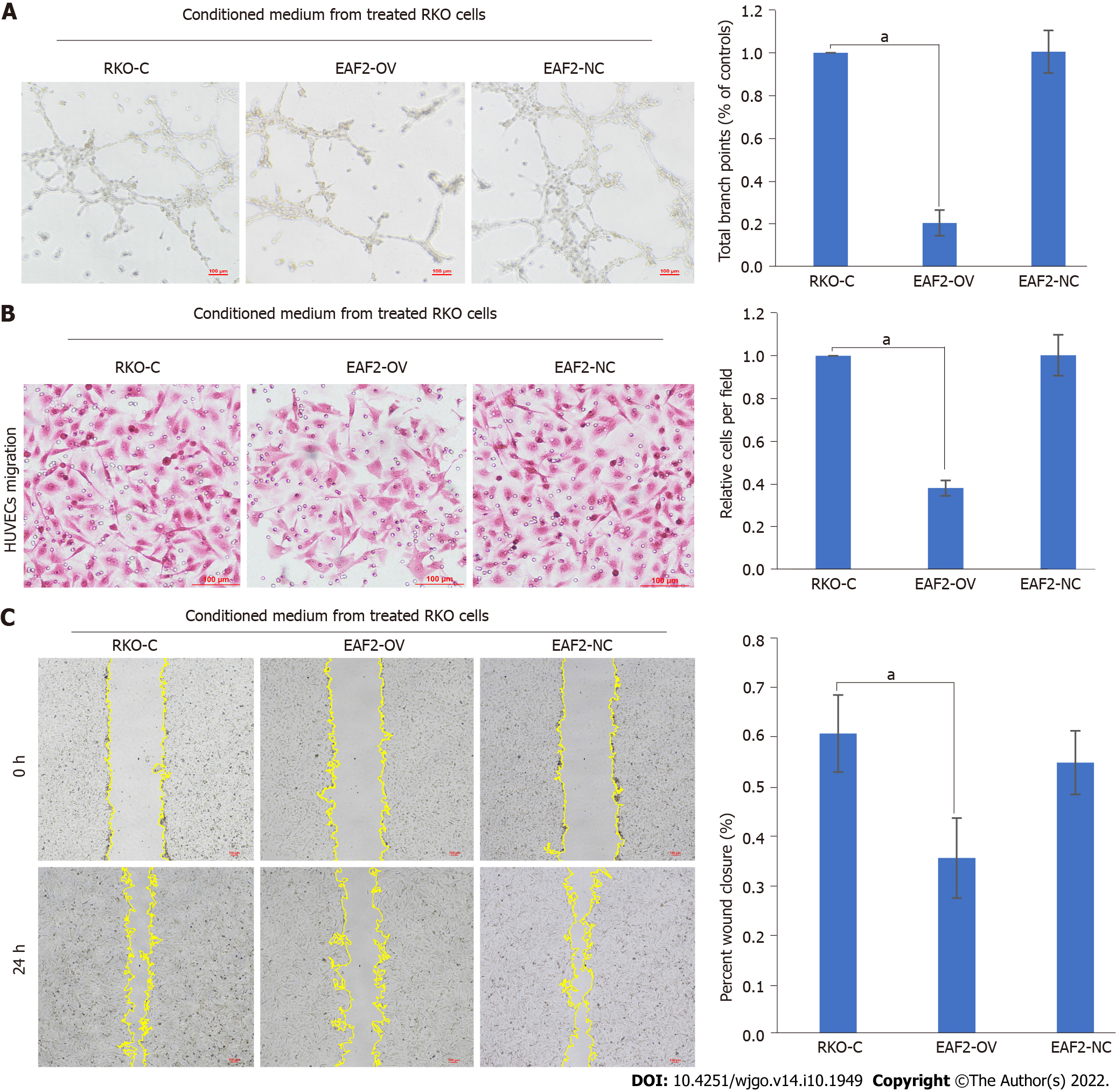
Figure 7 Overexpression of ELL-associated factor 2 in RKO cells inhibits tumor-induced angiogenesis.
A: Conditioned medium from RKO cells transfected with ELL-associated factor 2-overexpressed plasmids or empty control plasmids was collected to perform tube formation assay; B: Migration ability of human umbilical vein endothelial cells (HUVECs) analyzed by transwell migration assay; C: Migration ability of HUVECs analyzed by wound healing assay. And the results were measured with ImageJ software. The data are shown as the mean ± SD. aP < 0.001. HUVECs: Human Umbilical Vein Endothelial Cells; RKO-C: The group of Human Umbilical Vein Endothelial Cells cultured with conditioned medium from RKO cells cultured with 10% serum for 24 h; EAF2-OV: The group of human umbilical vein endothelial cells cultured with conditioned medium from RKO cells transfected with ELL-associated factor 2-overexpressed plasmids for 24 h; EAF2-NC: The group of human umbilical vein endothelial cells cultured with conditioned medium from RKO cells transfected with empty control plasmids for 24 h.
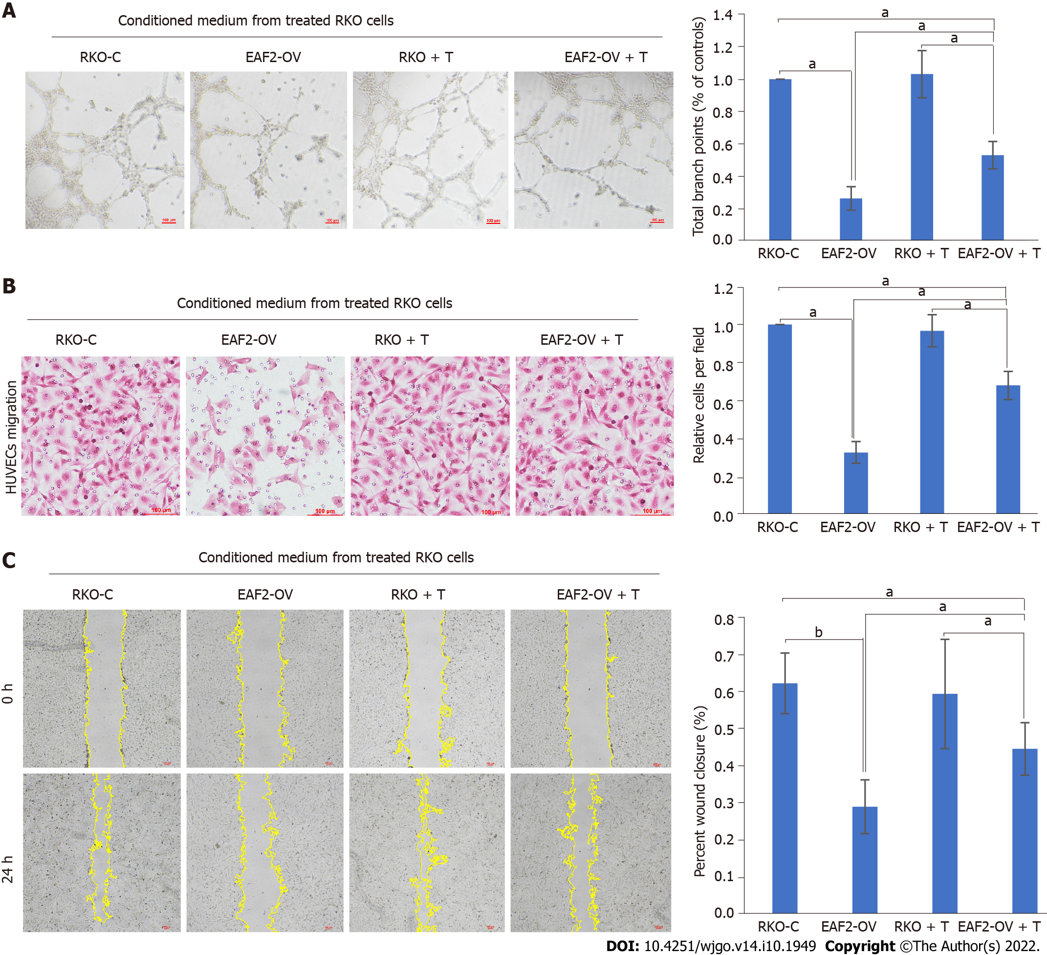
Figure 8 Overexpression of ELL-associated factor 2 inhibited tumor-induced angiogenesis in RKO cells via the signal transducer and activator of transcription 3/transforming growth factor beta 1 pathway.
A: Conditioned medium from RKO cells transfected with ELL-associated factor 2-overexpressed plasmids or treated with transforming growth factor beta 1 recombinant protein was collected to perform tube formation assay; B: Migration ability of human umbilical vein endothelial cells (HUVECs) analyzed by transwell migration assay; C: Migration ability of HUVECs analyzed by wound healing assay. And the results were measured with ImageJ software. The data are shown as the mean ± SD. aP < 0.05, bP < 0.001. RKO-C: The group of human umbilical vein endothelial cells cultured with conditioned medium from RKO cells cultured with 10% serum for 24 h; EAF2-OV: The group of human umbilical vein endothelial cells cultured with conditioned medium from RKO cells transfected with ELL-associated factor 2-overexpressed plasmids for 24 h; RKO + T: The group of human umbilical vein endothelial cells cultured with conditioned medium from RKO cells treated with 5 ng/mL transforming growth factor-β1 pathway recombinant protein for 24 h; EAF2-OV + T: The group of human umbilical vein endothelial cells cultured with conditioned medium from RKO cells which were transfected with EAF-OV plasmid for 24 h and then cultured with 5 ng/mL transforming growth factor beta 1 pathway recombinant protein for 24 h.
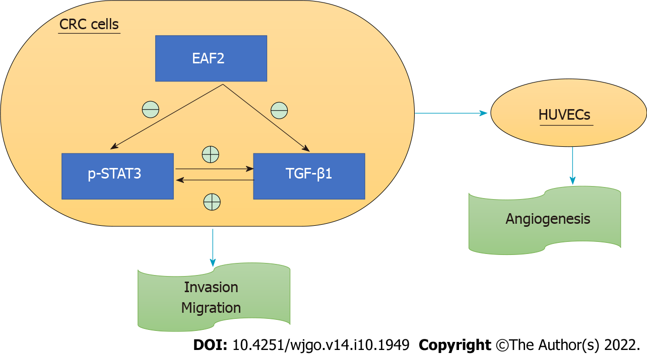
Figure 9 Schematic depiction of ELL-associated factor 2 regulating the signal transducer and activator of transcription 3/transforming growth factor beta 1 crosstalk pathway in this study.
EAF2: ELL-associated factor 2; p-STAT3: Phosphorylated-signal transducer and activator of transcription 3; TGF-β1: Transforming growth factor beta 1; CRC: Colorectal cancer; HUVECs: Human umbilical vein endothelial cells.
- Citation: Feng ML, Wu C, Zhang HJ, Zhou H, Jiao TW, Liu MY, Sun MJ. Overexpression of ELL-associated factor 2 suppresses invasion, migration, and angiogenesis in colorectal cancer. World J Gastrointest Oncol 2022; 14(10): 1949-1967
- URL: https://www.wjgnet.com/1948-5204/full/v14/i10/1949.htm
- DOI: https://dx.doi.org/10.4251/wjgo.v14.i10.1949


