Copyright
©The Author(s) 2022.
World J Gastrointest Oncol. Jan 15, 2022; 14(1): 153-162
Published online Jan 15, 2022. doi: 10.4251/wjgo.v14.i1.153
Published online Jan 15, 2022. doi: 10.4251/wjgo.v14.i1.153
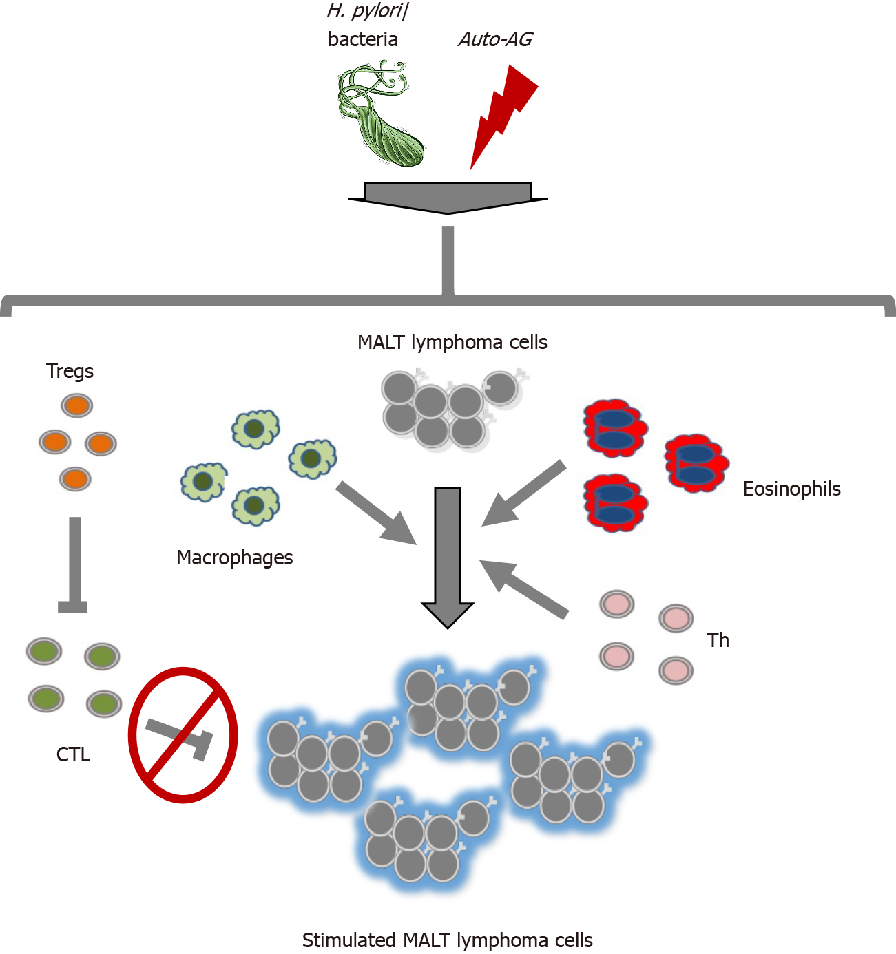
Figure 1 Graphical depiction of the interplay of mucosa-associated lymphoid tissue lymphoma cells with their microenvironment.
Helicobacter pylori (H. pylori), other bacteria and/or autoantigens (auto-AGs) support an immune regulatory microenvironment promoting mucosa-associated lymphoid tissue (MALT) lymphomagenesis in different organs. First, regulatory T cells are activated and suppress the immune response by maintaining H. pylori colonialization and influencing cytotoxic T cells, which possess malfunctions and therefore cannot inhibit the expansion of MALT lymphoma cells. Second, eosinophils and macrophages express a proliferation-inducing ligand (APRIL) and B cell-activating factor, supporting lymphomagenesis. The production of APRIL is induced by H. pylori antigens and H. pylori-specific T cells. Third, T helper cells and their cytokines (IL-4, Il-5, and IL-10) promote the growth and differentiation of lymphoma cells and are stimulated by H. pylori and/or auto-AGs. MALT: Mucosa-associated lymphoid tissue; H. pylori: Helicobacter pylori; Tregs: Regulatory T cells; CTL: Cytotoxic T cell; Auto-AG: Autoantigen; Th: T helper.
- Citation: Uhl B, Prochazka KT, Fechter K, Pansy K, Greinix HT, Neumeister P, Deutsch AJ. Impact of the microenvironment on the pathogenesis of mucosa-associated lymphoid tissue lymphomas. World J Gastrointest Oncol 2022; 14(1): 153-162
- URL: https://www.wjgnet.com/1948-5204/full/v14/i1/153.htm
- DOI: https://dx.doi.org/10.4251/wjgo.v14.i1.153


