Copyright
©The Author(s) 2021.
World J Gastrointest Oncol. Jul 15, 2021; 13(7): 662-672
Published online Jul 15, 2021. doi: 10.4251/wjgo.v13.i7.662
Published online Jul 15, 2021. doi: 10.4251/wjgo.v13.i7.662
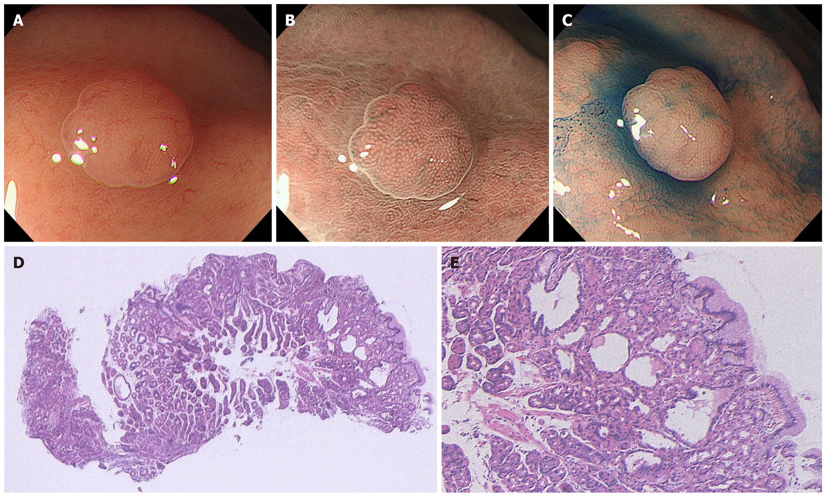
Figure 1 A case of sporadic fundic gland polyp without dysplasia.
A: White light endoscopic view. A 47-year-old man with a complaint of heartburn underwent esophagogastroduodenoscopy. He had no medical history of receiving long-term proton pump inhibitor therapy and no family history of familial adenomatous polyposis (FAP). White light endoscopy identified several fundic gland polyp-like lesions in the body of the stomach without atrophic mucosa, suggesting a Helicobacter pylori-uninfected setting. Among them, a 3 mm isochromatic sessile polyp with a smooth surface structure was detected in the middle body; B: Magnifying narrow-band imaging (NBI) endoscopic view. Magnifying NBI endoscopy revealed regularly arranged, white, dot-shaped crypt openings resembling surrounding mucosa; C: Chromoendoscopic view. Chromoendoscopy using indigo carmine dye revealed less irregularity of the lesion surface. This lesion was endoscopically diagnosed as a fundic gland polyp without dysplasia and was biopsied; D and E: Histopathological view. The histopathological examination revealed a non-dysplastic fundic gland polyp with hyperplastic proliferation of the oxyntic glands and several cystically dilated glands. Colonoscopy did not reveal indications for FAP.
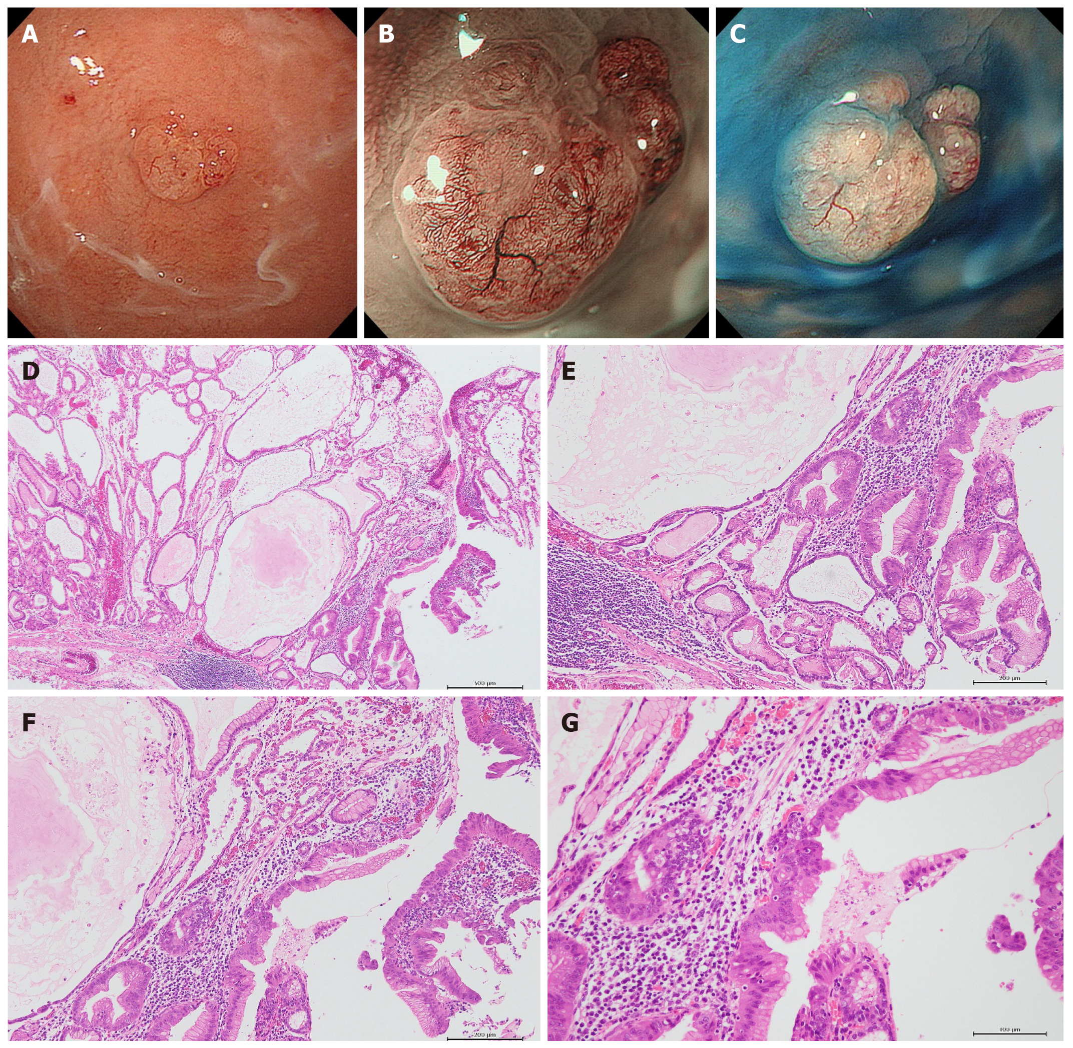
Figure 2 A case of sporadic fundic gland polyp with carcinoma.
A: White light endoscopic view. A 73-year-old woman with a complaint of upper abdominal discomfort underwent esophagogastroduodenoscopy. She had received long-term proton pump inhibitor (PPI) therapy for gastroesophageal reflux disease. White light endoscopy identified several fundic gland polyp-like lesions in the body and fundus of the stomach without atrophic mucosa, suggesting a Helicobacter pylori (H. pylori)-uninfected setting. Among them, a 6 mm isochromatic sessile polyp with a slightly irregular surface structure was detected in the fundus; B: Magnifying narrow-band imaging (NBI) endoscopic view. Magnifying NBI endoscopy revealed irregular microvessels on the lesion surface; C: Chromoendoscopic view. Chromoendoscopy using indigo carmine dye more clearly revealed the irregularity of the lesion surface. This lesion was endoscopically diagnosed as a fundic gland polyp with dysplasia or carcinoma and was removed by the endoscopic mucosal resection technique; D: Histopathological view (low magnification). The histopathological examination revealed a fundic gland polyp with prominent dilated cystic glands often observed in patients receiving PPI therapy; E-G: Histopathological view (higher magnification of D). This lesion coexisted with epithelial dysplasia and intramucosal well-differentiated adenocarcinoma. Serological examination confirmed that she was negative for H. pylori infection. Colonoscopy as well as her family history showed no familial adenomatous polyposis.
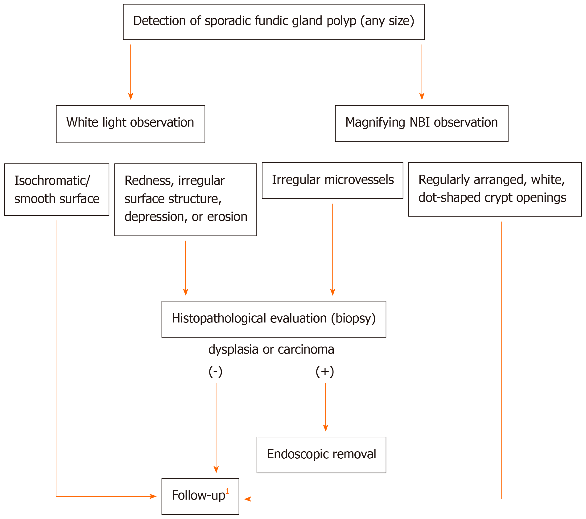
Figure 3 Proposal of a management algorithm for sporadic fundic gland polyps.
1If multiple fundic gland polyps are detected in patients receiving proton pump inhibitor therapy, reducing or discontinuing proton pump inhibitor therapy or switching to H2-receptor antagonists is recommended. NBI: Narrow-band imaging.
- Citation: Sano W, Inoue F, Hirata D, Iwatate M, Hattori S, Fujita M, Sano Y. Sporadic fundic gland polyps with dysplasia or carcinoma: Clinical and endoscopic characteristics. World J Gastrointest Oncol 2021; 13(7): 662-672
- URL: https://www.wjgnet.com/1948-5204/full/v13/i7/662.htm
- DOI: https://dx.doi.org/10.4251/wjgo.v13.i7.662


