Copyright
©The Author(s) 2020.
World J Gastrointest Oncol. Sep 15, 2020; 12(9): 1073-1079
Published online Sep 15, 2020. doi: 10.4251/wjgo.v12.i9.1073
Published online Sep 15, 2020. doi: 10.4251/wjgo.v12.i9.1073
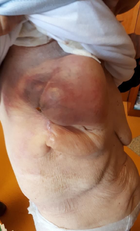
Figure 1 Mass of 10 cm × 5 cm on the right breast and lymphadenopathy in the right axillary cable of about 8.
5 cm at palpation.
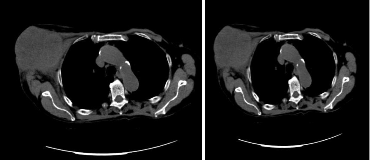
Figure 2 Computed tomography scan revealed coarse swelling of 11 cm × 6 cm at the right mammary level, ascending colon lesion and 3 cm lymphadenopathy.
No obvious lesions on other parenchyma.
Figure 3 Microscopy examination of the breast specimen.
A: Hematoxylin-Eosin: Adenocarcinoma with cribriform pattern (200 ×); B: Intense nuclear staining of the neoplastic cells for CDX2 at immunohistochemistry (× 400); and C: Microscopy examination of the breast specimen.
Figure 4 Microscopy examination of the colon specimen.
A: Clusters of poorly differentiated neoplastic cells in solid and cribriform pattern and desmoplastic stromal reaction (× 100); B: Area with abundant extracellular mucin (left) (× 100); and C: CDX2 showed diffuse, nuclear positive staining in neoplastic cells (× 200).
Figure 5 Breast biopsy.
A: Invasive adenocarcinoma; B: A focal cribriform pattern, necrosis area, brisk mitotic activity and muscular infiltration (arrow) (Hematoxylin-Eosin, 100 ×, 200 ×); and C: A focal cribriform pattern, necrosis area, brisk mitotic activity and muscular infiltration (arrow) (Hematoxylin-Eosin, 200 ×).
- Citation: Taccogna S, Gozzi E, Rossi L, Caruso D, Conte D, Trenta P, Leoni V, Tomao S, Raimondi L, Angelini F. Colorectal cancer metastatic to the breast: A case report. World J Gastrointest Oncol 2020; 12(9): 1073-1079
- URL: https://www.wjgnet.com/1948-5204/full/v12/i9/1073.htm
- DOI: https://dx.doi.org/10.4251/wjgo.v12.i9.1073


