Copyright
©The Author(s) 2020.
World J Gastrointest Oncol. Sep 15, 2020; 12(9): 1031-1043
Published online Sep 15, 2020. doi: 10.4251/wjgo.v12.i9.1031
Published online Sep 15, 2020. doi: 10.4251/wjgo.v12.i9.1031
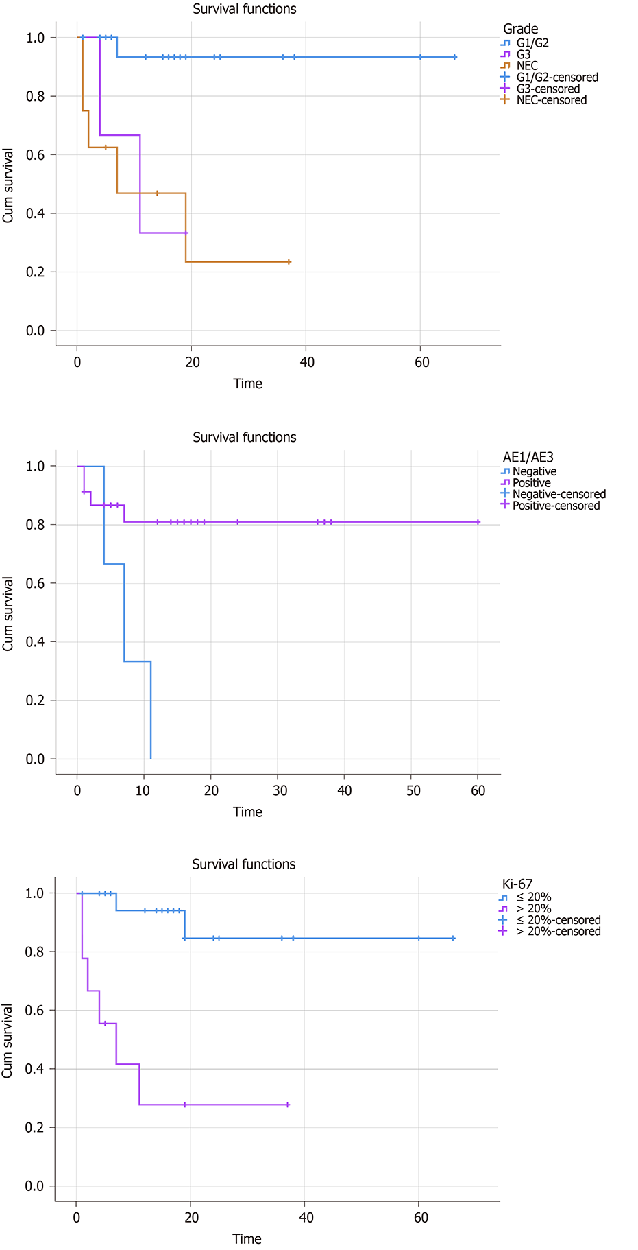
Figure 1 Survival analysis showed that tumor grade, AE1/AE3, and Ki-67 were significantly related to the overall survival rate.
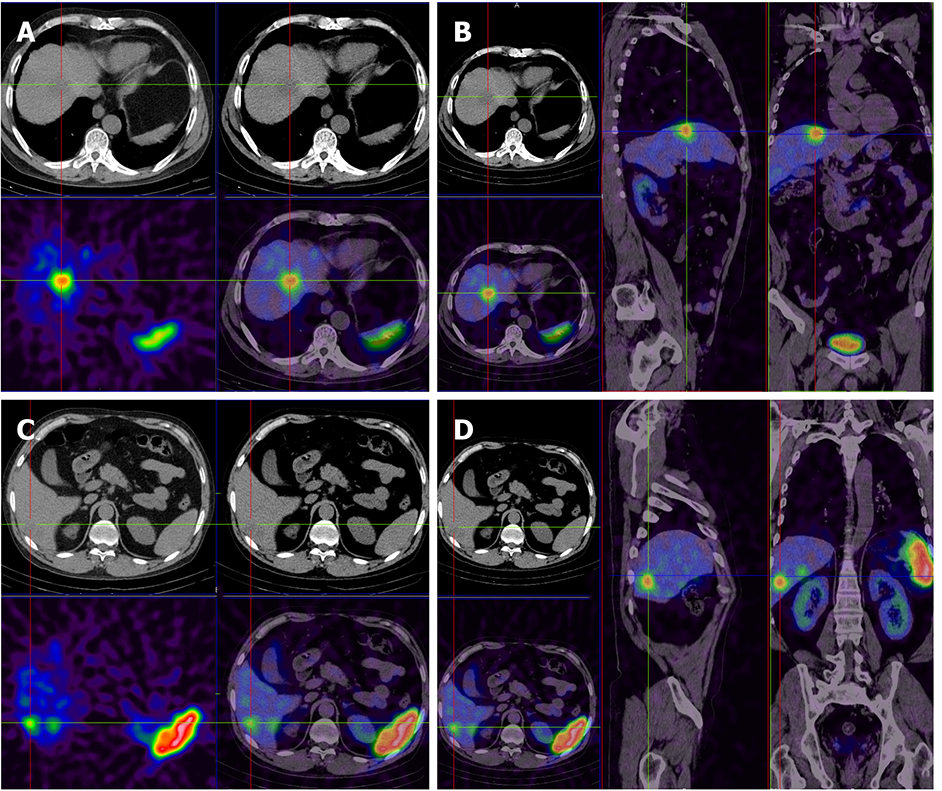
Figure 2 There are multiple low-density lesions in the liver with nonuniform radioactive distribution.
A and B: The size of the largest lesion is 2.3 cm × 2.8 cm, and the computed tomography value is 36 HU; C and D: Smaller lesion.
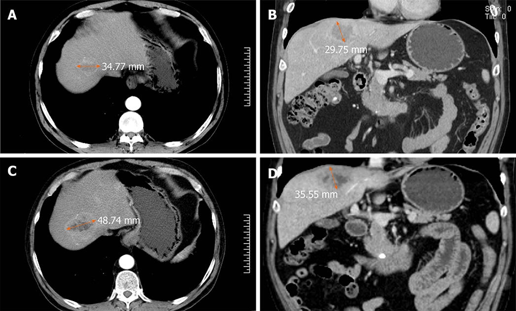
Figure 3 Comparison of computed tomography images of the liver lesion before and after long-acting repeatable octreotide treatment.
A and B: Pre-treatment images; C and D: Post-treatment images.
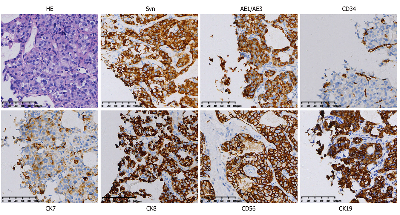
Figure 4 Hematoxylin-eosin staining indicated the possibility of cancer, and further immunohistochemistry was performed.
The tumor was regarded as a highly differentiated neuroendocrine tumor according to the results. The tumor was positive for Syn, AE1/AE3, CD34, CK7, CK8, CD56, and CK19 (magnification, 200 ×).
Figure 5 Positron emission tomography-computed tomography was carried out after surgery and showed that the margin of the low-density shadow in the right hepatic lobe was more active and more likely attributable to postoperative changes.
No other obvious abnormalities were observed.
- Citation: Wang HH, Liu ZC, Zhang G, Li LH, Li L, Meng QB, Wang PJ, Shen DQ, Dang XW. Clinical characteristics and outcome of primary hepatic neuroendocrine tumors after comprehensive therapy. World J Gastrointest Oncol 2020; 12(9): 1031-1043
- URL: https://www.wjgnet.com/1948-5204/full/v12/i9/1031.htm
- DOI: https://dx.doi.org/10.4251/wjgo.v12.i9.1031


