Copyright
©The Author(s) 2020.
World J Gastrointest Oncol. Jun 15, 2020; 12(6): 663-676
Published online Jun 15, 2020. doi: 10.4251/wjgo.v12.i6.663
Published online Jun 15, 2020. doi: 10.4251/wjgo.v12.i6.663
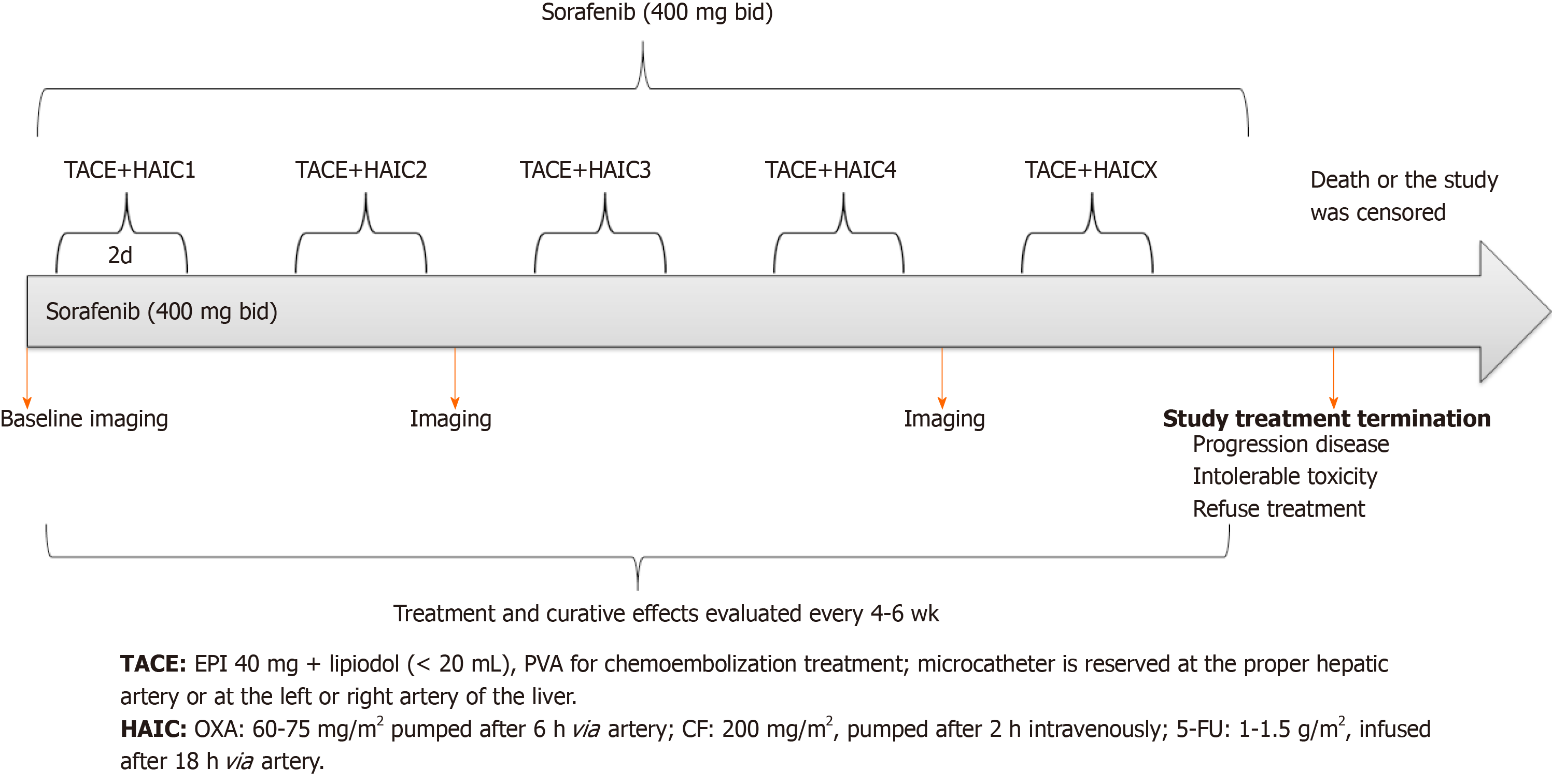
Figure 1 Scheme of transcatheter arterial chemoembolization-hepatic arterial infusion chemotherapy and sorafenib.
TACE: Transcatheter arterial chemoembolization; HAIC: Hepatic arterial infusion chemotherapy; 5-FU: 5-fluorouracil; PVA: Polyvinyl alcohol; CF: Calcium folinate; EPI: Epirubicin; OXA: Oxaliplatin.
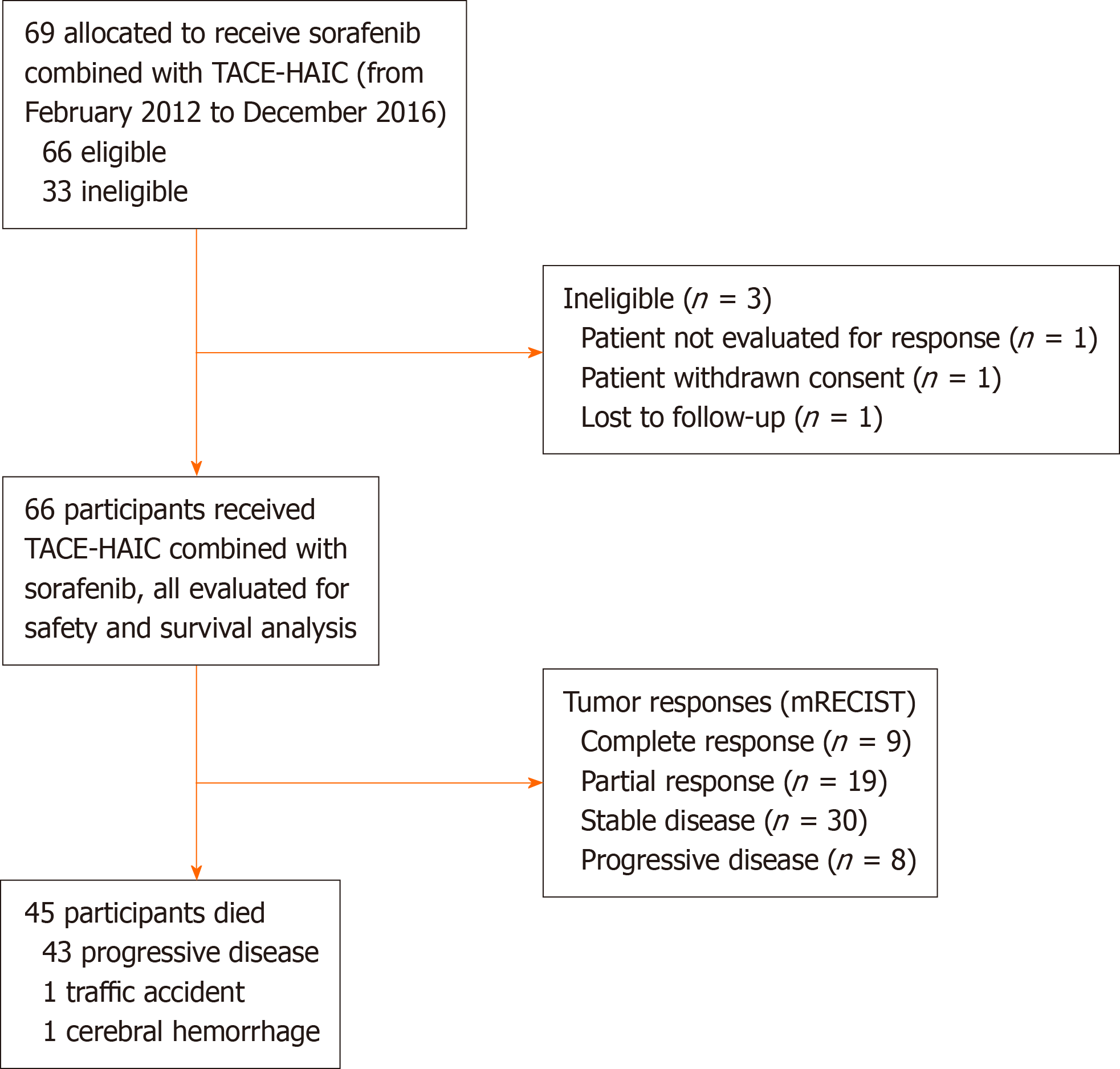
Figure 2 Patient selection flowchart.
mRECIST: Modified Response Evaluation Criteria in Solid Tumors; TACE: Transarterial chemoembolization; HAIC: Hepatic arterial infusion chemotherapy.
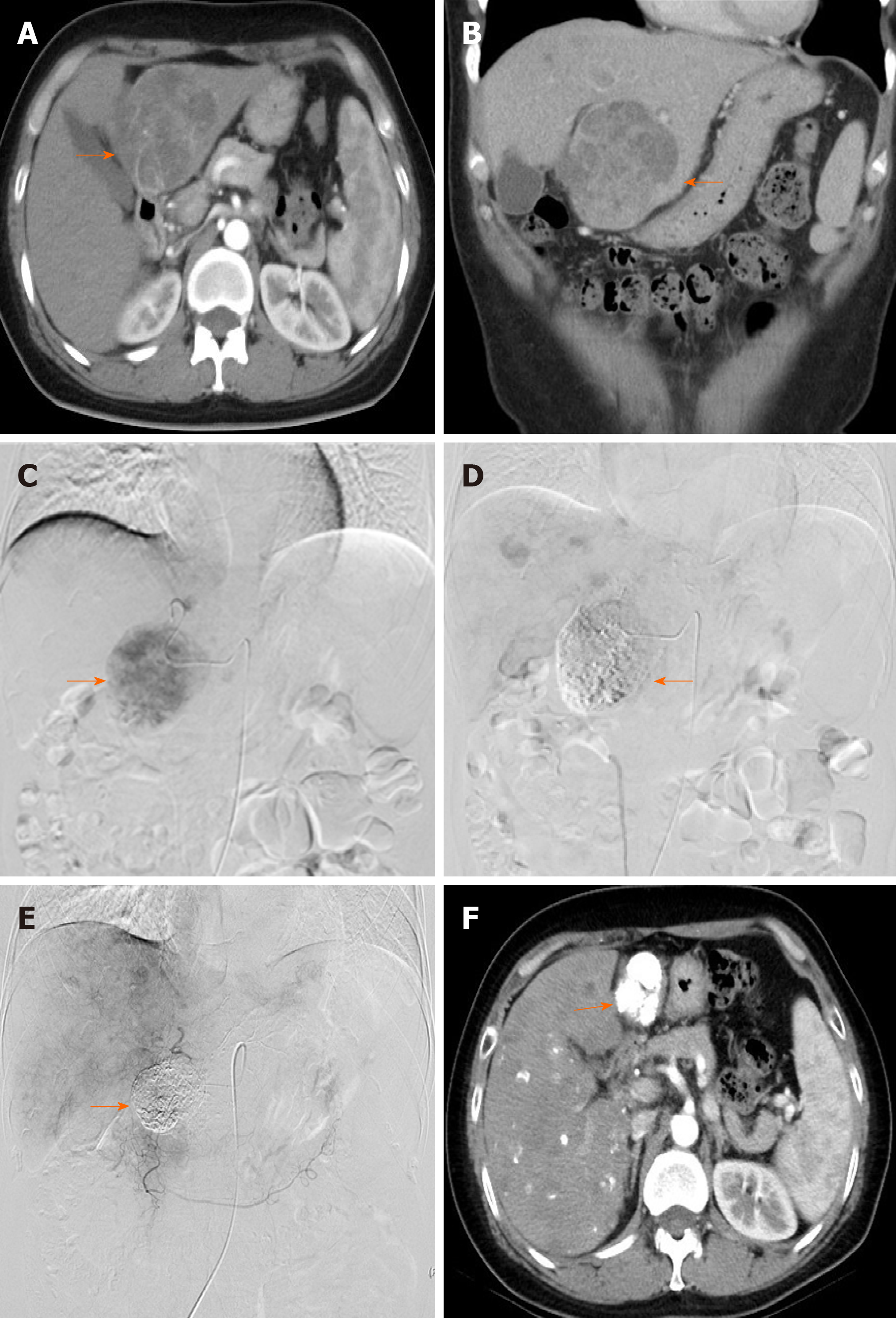
Figure 3 Images from a 41-year-old woman with intermediate-stage hepatocellular carcinoma who underwent combination therapy with sorafenib with hepatic arterial infusion chemotherapy (folinic acid, 5-fluorouracil, and oxaliplatin) after transarterial chemoembolization.
A and B: Preoperative computed tomography (CT) showed the lesion in the liver (arrow); C: Angiography via the proper hepatic artery revealed a tumor mass in the liver (arrow); D: Postoperative angiography revealed the scattered deposition of lipiodol within the lesions in the liver (arrow); E: After 1 year, angiography via the proper hepatic artery revealed scattered deposition of lipiodol within the lesions in the liver (arrow); F: After 1 year, CT revealed scattered deposition of lipiodol within the lesions in the liver, and the tumor lesions had no enhancement during the arterial phase (arrow).
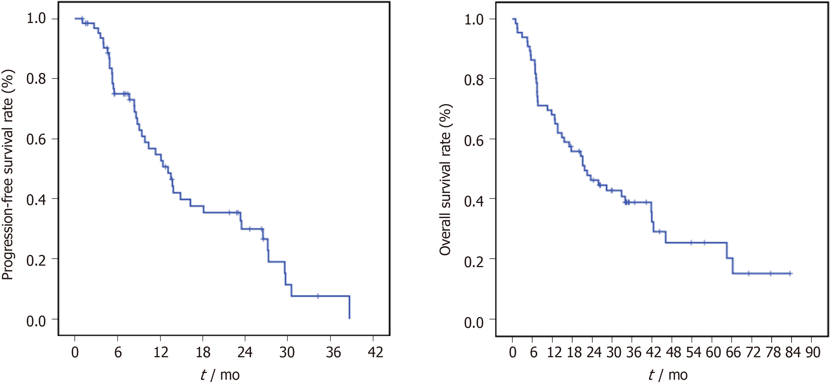
Figure 4 Kaplan-Meier curves of the progression-free survival and overall survival of patients with hepatocellular carcinoma who were treated with sorafenib combined with hepatic arterial infusion chemotherapy (folinic acid, 5-fluorouracil, and oxaliplatin) after transarterial chemoembolization and for Barcelona Clinic Liver Cancer stage B/C hepatocellular carcinoma.
- Citation: Liu BJ, Gao S, Zhu X, Guo JH, Zhang X, Chen H, Wang XD, Yang RJ. Sorafenib combined with embolization plus hepatic arterial infusion chemotherapy for inoperable hepatocellular carcinoma. World J Gastrointest Oncol 2020; 12(6): 663-676
- URL: https://www.wjgnet.com/1948-5204/full/v12/i6/663.htm
- DOI: https://dx.doi.org/10.4251/wjgo.v12.i6.663


