Copyright
©The Author(s) 2020.
World J Gastrointest Oncol. Apr 15, 2020; 12(4): 435-446
Published online Apr 15, 2020. doi: 10.4251/wjgo.v12.i4.435
Published online Apr 15, 2020. doi: 10.4251/wjgo.v12.i4.435
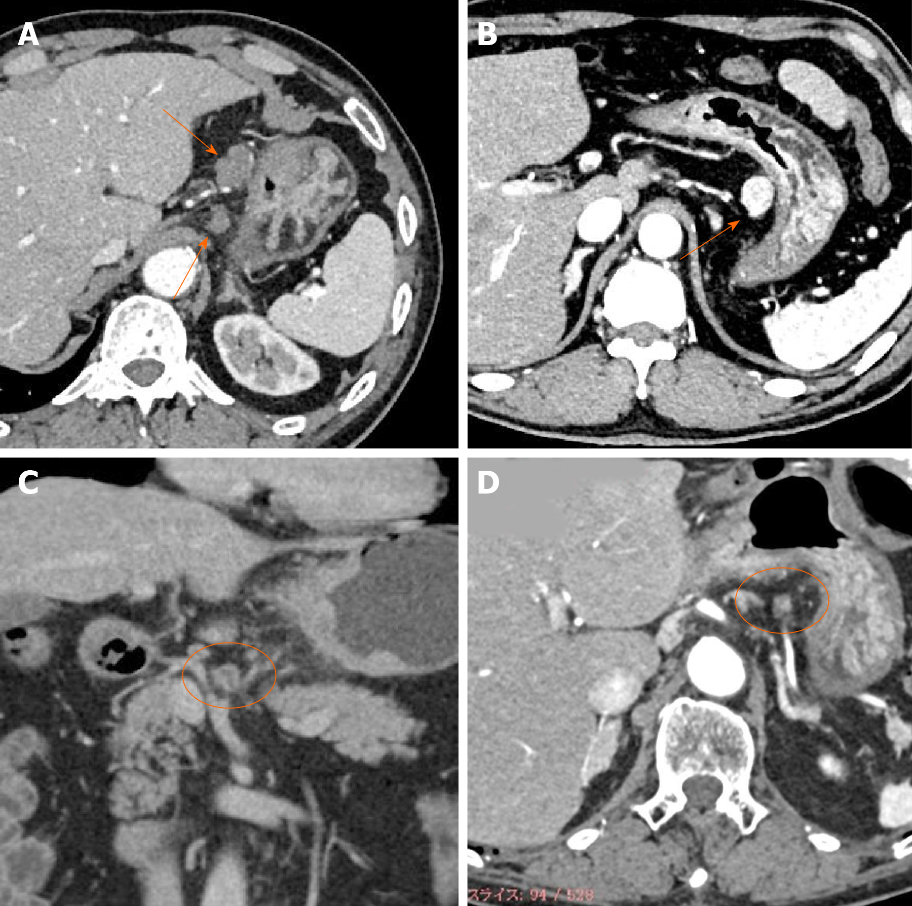
Figure 1 Typical images of four types of enhancement of metastatic lymph nodes.
A: Weak enhancement; B: Obvious enhancement; C: Ring enhancement; D: Partial enhancement.
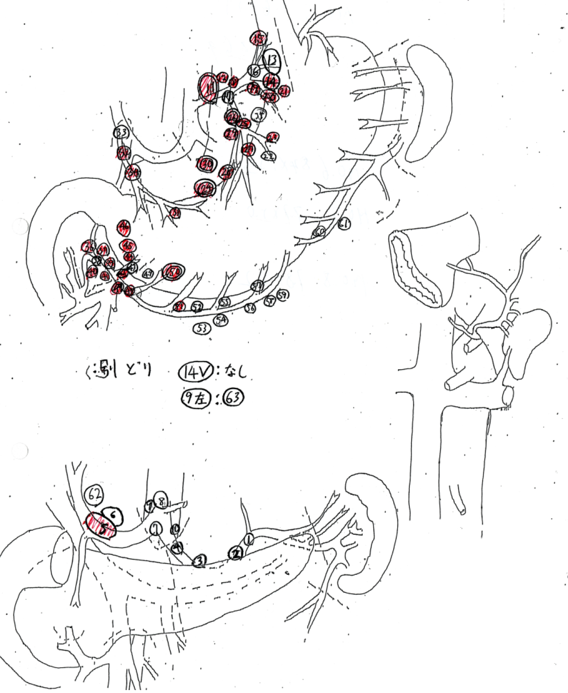
Figure 2 Sample of a lymph node map routinely created in our hospital.
The dissected lymph nodes were harvested, separated individually, and the location of the nodes recorded with serial numbers on a map. In the prospective study, sizes of each lymph node were also measured. Red markers on maps are pathologically confirmed metastatic lymph nodes.
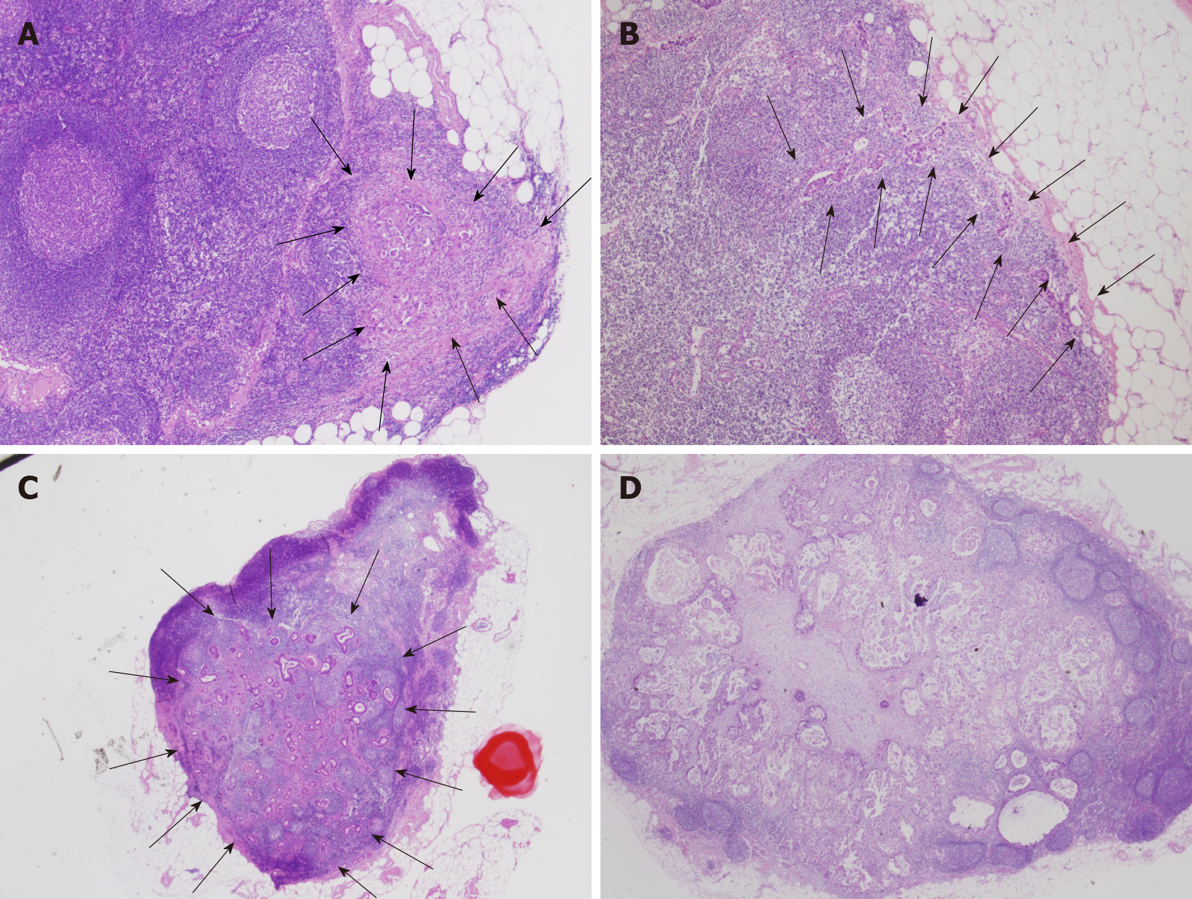
Figure 3 Typical images of intranodal morphological metastatic patterns.
A: Small nodular type; B: Peripheral type; C: Large nodular type; D: Diffuse type.
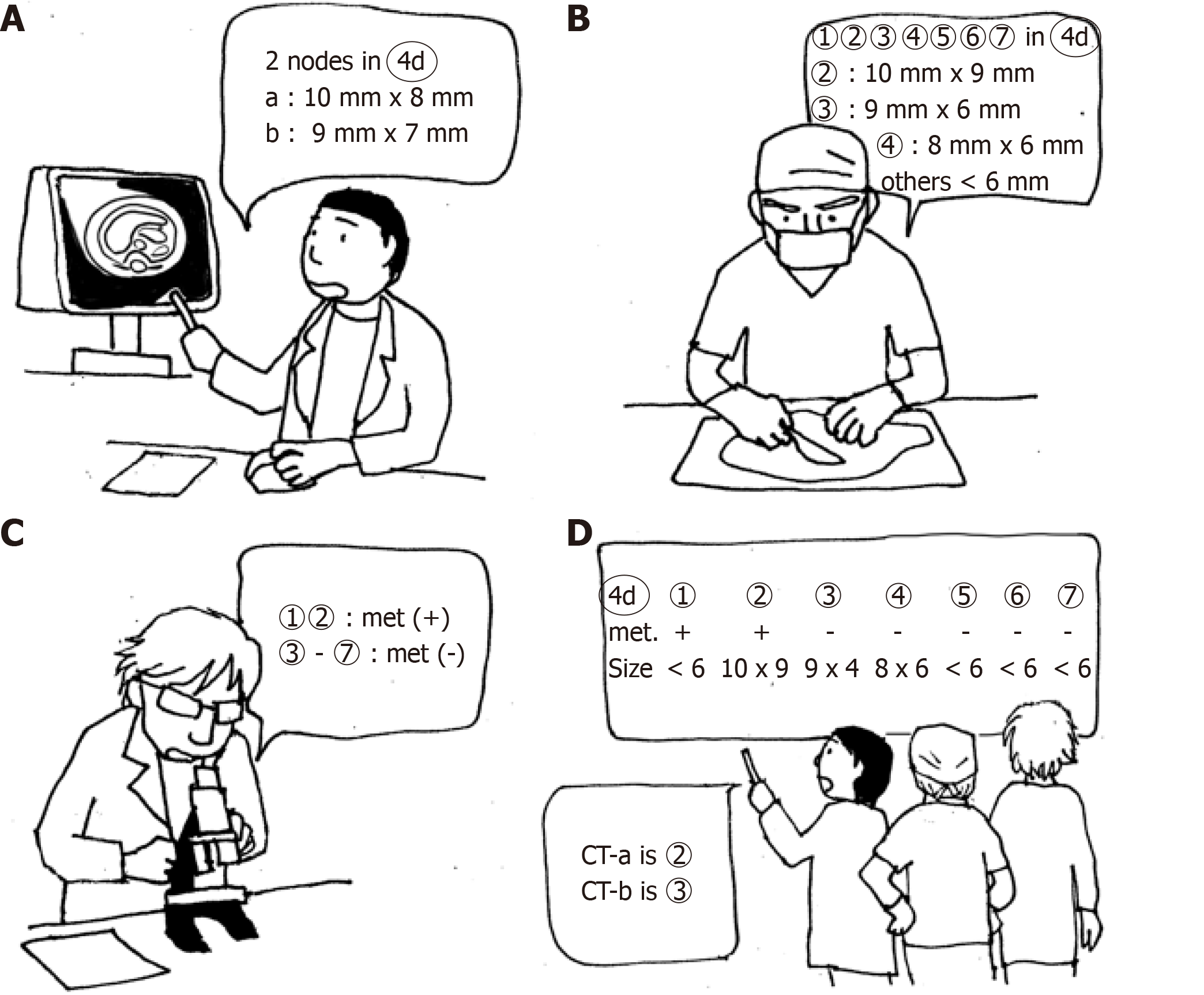
Figure 4 A schema for precise one-to-one correspondence of lymph nodes.
A: CT examination before surgery. B: Lymph node mapping. All dissected nodes were mapped and measured when the size was > 7 mm. C: Pathological examination. D: One-to-one correspondence between CT images and pathological results by using the size data of the map.
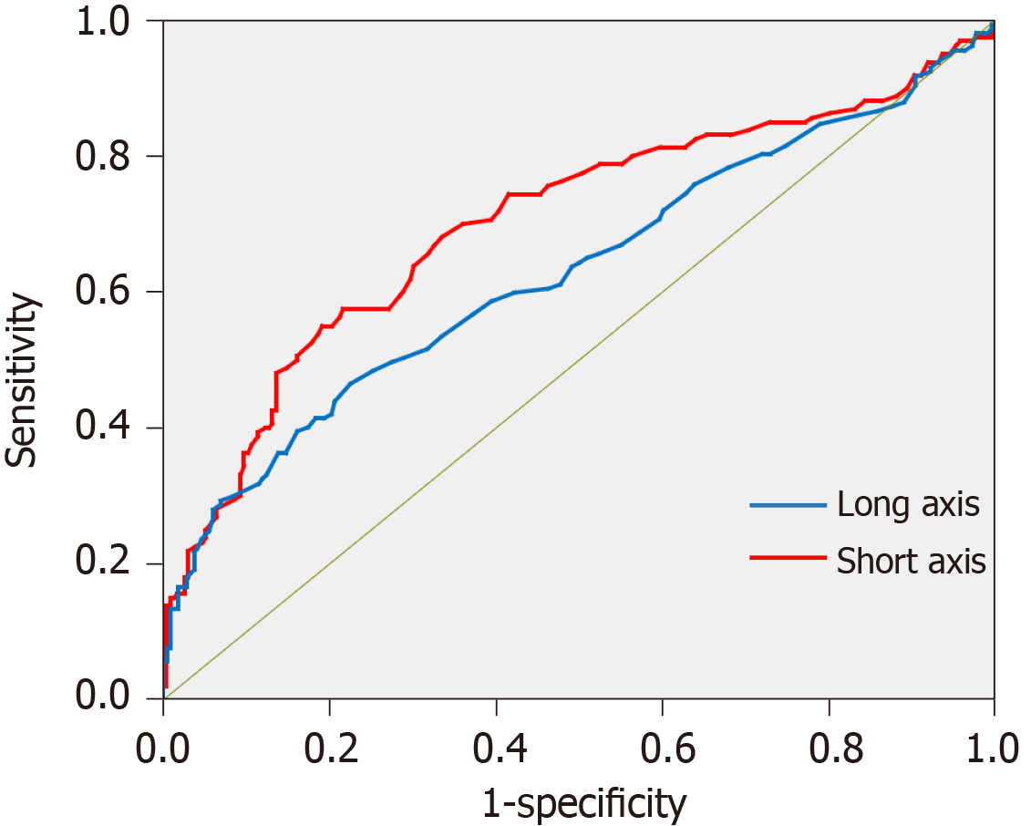
Figure 5 Receiver-operating characteristic curve for diagnosis of nodal metastasis using multi-detector computed tomography created from the data of the retrospective study.
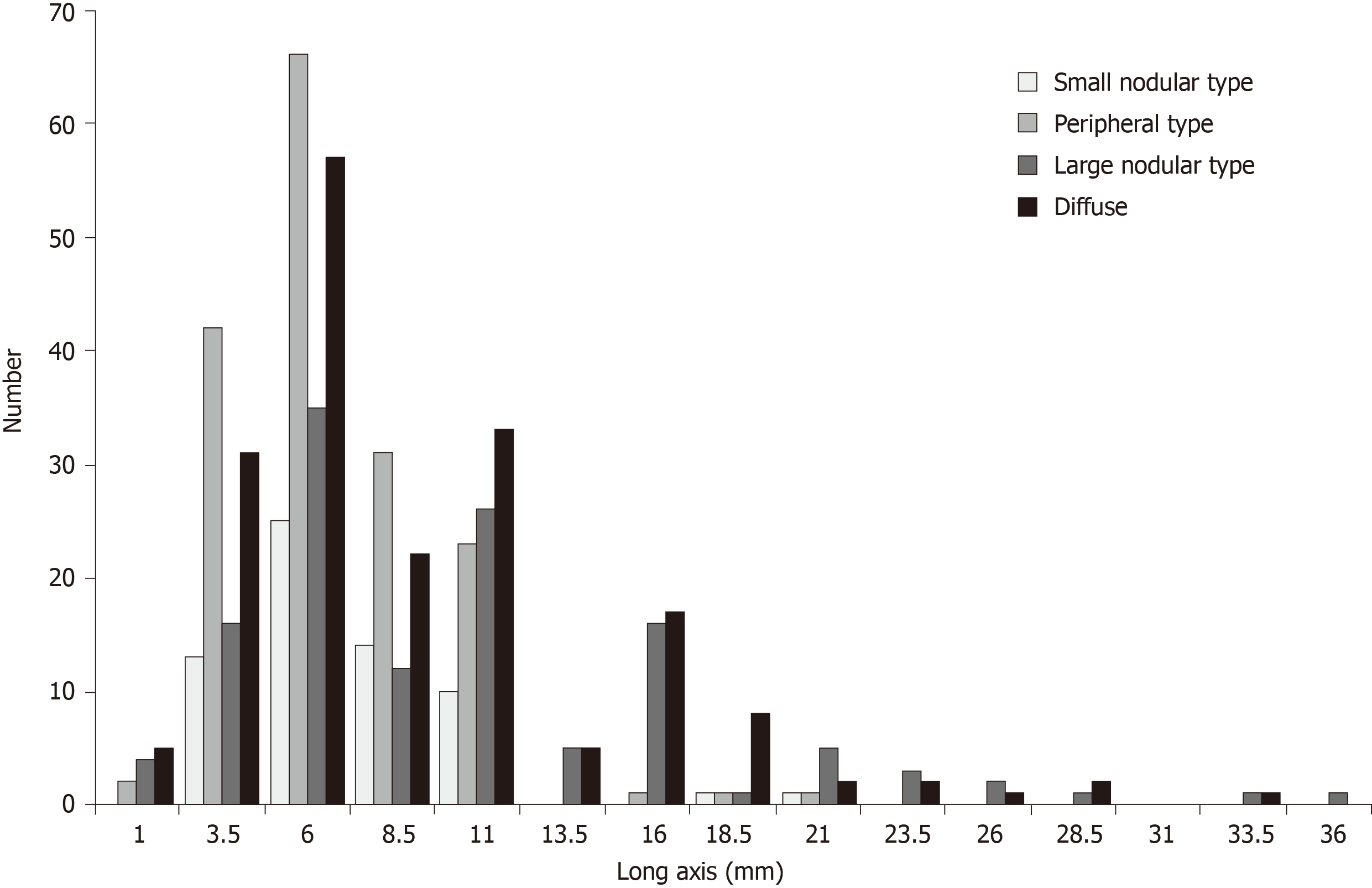
Figure 6 Distribution of lymph node metastasis in four types based on the long axis.
- Citation: Jiang ZY, Kinami S, Nakamura N, Miyata T, Fujita H, Takamura H, Ueda N, Kosaka T. Diagnostic ability of multi-detector spiral computed tomography for pathological lymph node metastasis of advanced gastric cancer. World J Gastrointest Oncol 2020; 12(4): 435-446
- URL: https://www.wjgnet.com/1948-5204/full/v12/i4/435.htm
- DOI: https://dx.doi.org/10.4251/wjgo.v12.i4.435


