Copyright
©The Author(s) 2020.
World J Gastrointest Oncol. Oct 15, 2020; 12(10): 1209-1215
Published online Oct 15, 2020. doi: 10.4251/wjgo.v12.i10.1209
Published online Oct 15, 2020. doi: 10.4251/wjgo.v12.i10.1209
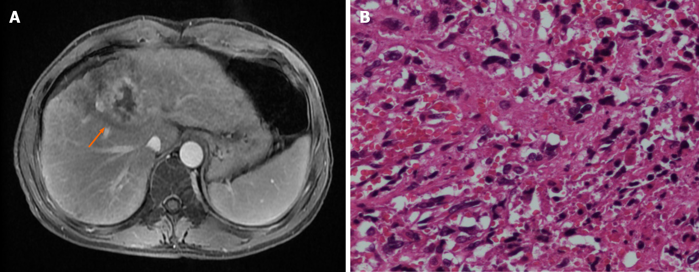
Figure 1 Magnetic resonance imaging and histology.
A: MRI showing the location of the tumor in the liver segments 4/8; B: Histology of the tumor tissue (40 ×). The tumor displayed fish-like changes throughout, with atypical cells and lymphocyte infiltration observed.
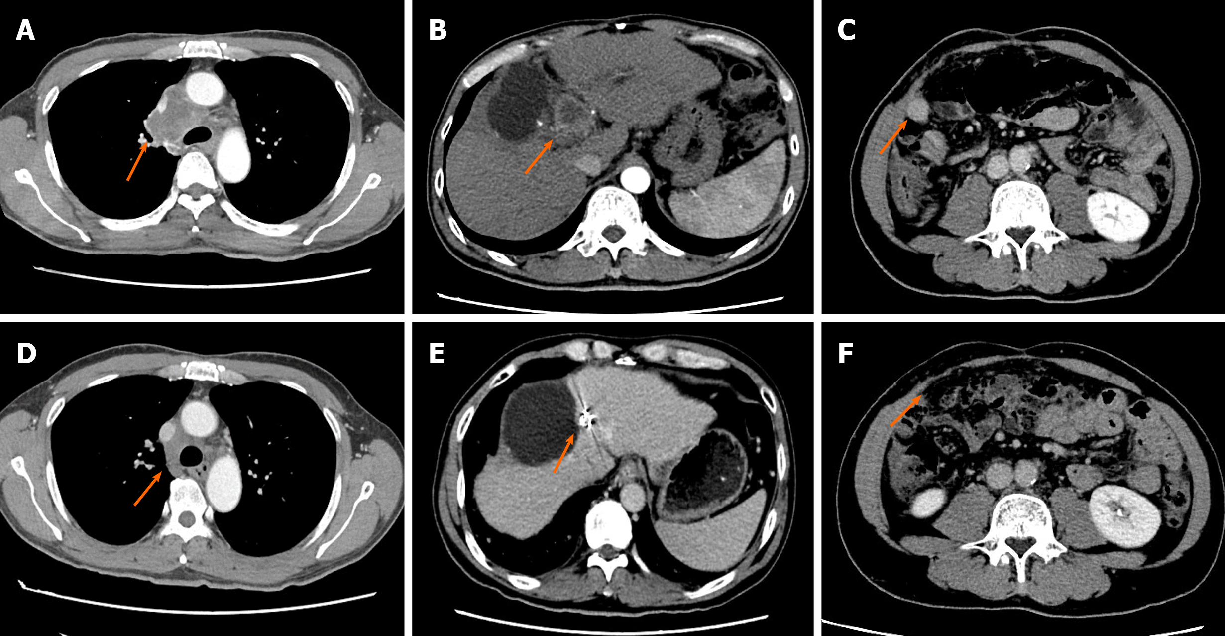
Figure 2 Computed tomography scans before and after immunotherapy.
A-C: Computed tomography scan showing metastasis of the mediastinal lymph nodes, recurrent hepatic lesions that led to compression of the bile duct, and a new metastatic lesion in the abdominal cavity; D-F: Metastatic lesions showing achievement of complete response2 mo after receiving nivolumab. In addition, compression of the bile duct was relieved.
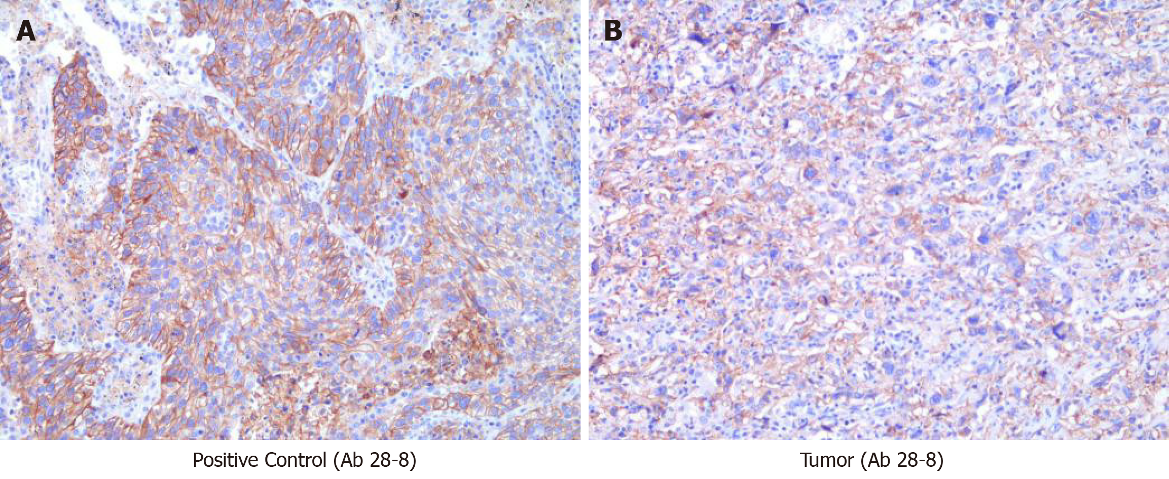
Figure 3 Immunohistochemical staining for programmed death-ligand 1 (40 ×).
A: Positive control (Ab 28-8); B: Tumor (Ab 28-8).
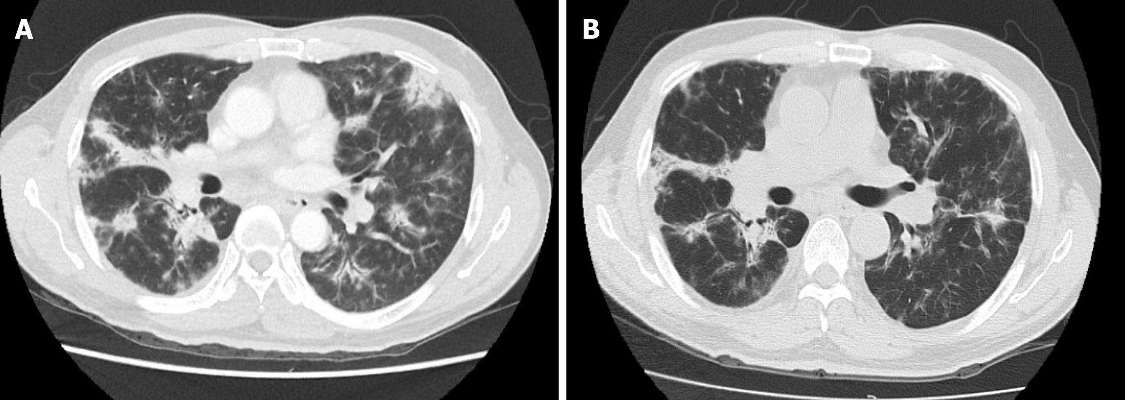
Figure 4 Computed tomography scans before and after corticosteroid treatment for interstitial pneumonia.
A: Computed tomography (CT) scan showing bilateral interstitial pneumonia caused by the PD-1 inhibitor; B: CT scan showing that pneumonia was alleviated by corticosteroid treatment.
- Citation: Zhu SG, Li HB, Yuan ZN, Liu W, Yang Q, Cheng Y, Wang WJ, Wang GY, Li H. Achievement of complete response to nivolumab in a patient with advanced sarcomatoid hepatocellular carcinoma: A case report. World J Gastrointest Oncol 2020; 12(10): 1209-1215
- URL: https://www.wjgnet.com/1948-5204/full/v12/i10/1209.htm
- DOI: https://dx.doi.org/10.4251/wjgo.v12.i10.1209


